. 2011 Sep 14;31(37):13272-80.
doi: 10.1523/JNEUROSCI.3337-11.2011.

Adenosine receptor signaling modulates permeability of the blood-brain barrier

Affiliations

Adenosine receptor signaling modulates permeability of the blood-brain barrier

Aaron J Carman et al. J Neurosci. .

Abstract

The blood-brain barrier (BBB) is comprised of specialized endothelial cells that form the capillary microvasculature of the CNS and is essential for brain function. It also poses the greatest impediment in the treatment of many CNS diseases because it commonly blocks entry of therapeutic compounds. Here we report that adenosine receptor (AR) signaling modulates BBB permeability in vivo. A(1) and A(2A) AR activation facilitated the entry of intravenously administered macromolecules, including large dextrans and antibodies to β-amyloid, into murine brains. Additionally, treatment with an FDA-approved selective A(2A) agonist, Lexiscan, also increased BBB permeability in murine models. These changes in BBB permeability are dose-dependent and temporally discrete. Transgenic mice lacking A(1) or A(2A) ARs showed diminished dextran entry into the brain after AR agonism. Following treatment with a broad-spectrum AR agonist, intravenously administered anti-β-amyloid antibody was observed to enter the CNS and bind β-amyloid plaques in a transgenic mouse model of Alzheimer's disease (AD). Selective AR activation resulted in cellular changes in vitro including decreased transendothelial electrical resistance, increased actinomyosin stress fiber formation, and alterations in tight junction molecules. These results suggest that AR signaling can be used to modulate BBB permeability in vivo to facilitate the entry of potentially therapeutic compounds into the CNS. AR signaling at brain endothelial cells represents a novel endogenous mechanism of modulating BBB permeability. We anticipate these results will aid in drug design, drug delivery and treatment options for neurological diseases such as AD, Parkinson's disease, multiple sclerosis and cancers of the CNS.


Figures

Figure 1.
Figure 1.
NECA treatment increases BBB permeability in a temporally discrete and reversible manner. A, B, Dose-dependent increases in 10 kDa (A) and 70 kDa (B) dextrans into WT mouse brain 3 h after intravenous administration of NECA or vehicle as measured by fluorimetry (10–15 animals per group). C, Extravasation time course of 10 kDa FITC-dextran into WT mouse brain when coadministered intravenously with NECA (0.08 mg/kg) or vehicle, as measured by fluorimetry (10–15 animals per group). D, Extravasation time course of 10 kDa Texas Red-dextran, administered intravenously 90 min before harvest times (as displayed), into WT mouse brain tissue after intravenous pretreatment (time = 0) with NECA (0.08 mg/kg) or vehicle, as measured by fluorimetry (3–5 animals per group). Data are splined scatter plots with scaled time on the x-axis. Experiments were performed at least twice. *p ≤ 0.05, significant differences (Student's t test) from vehicle. Data are mean ± SEM.
Figure 2.
Figure 2.
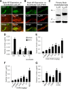
Increased BBB permeability depends on selective agonism of A1 and A2A ARs. A, B, Immunofluorescent staining (A) and fluorescence in situ hybridization (B) of CD31 (endothelial cell marker; green), and A1 (left column; red) and A2A (right column; red) ARs near the cortical area of the brain in naive mice. Scale bar, 20 μm. C, Western blot analysis of A1 (left) and A2A (right) AR expression in isolated primary BECs from naive mice. β-Actin expression is shown as a loading control. D, Decreased levels of dextran in brains of A1 and A2A AR knock-out mice 3 h after intravenous administration of NECA (0.08 mg/kg) or vehicle compared with WT mice, as measured by fluorimetry. No significant increase in dextran levels was detected in brains of A1 knock-out mice that were pretreated with the selective A2A antagonist SCH 58261 (5–8 animals per group). E, F, Dose-dependent entry of 10 kDa FITC-dextran into WT brain tissue 3 h after intravenous coadministration of CGS 21860 (selective A2A AR agonist) (E) or CCPA (selective A1 AR agonist) (F), as measured by fluorimetry. NECA (0.08 mg/kg) was used as a positive control (10–13 animals per group). G, Levels of 10 kDa FITC-dextran in WT mouse brain tissue 3 h after intravenous administration of vehicle, NECA (0.08 mg/kg), CGS 21680 (0.54 mg/kg), CCPA (0.37 mg/kg), and in combination [left column, CGS 21680 (0.54 mg/kg) + CCPA (0.37 mg/kg); right column CGS 21680 (0.54 mg/kg) + CCPA (0.037 mg/kg)] (3–4 mice per group). Experiments were repeated at least twice. *p ≤ 0.05, significant differences (Student's t test) from vehicle. Data are mean ± SEM.
Figure 3.
Figure 3.
The selective A2A AR agonist Lexiscan increases BBB permeability in murine models. A–D, Lexiscan administration increases BBB permeability to 10 kDa dextran in mice and rats. See Materials and Methods for experimental design. A, Data bars before the line break represent groups that received 3 Lexiscan injections. The bar after the line break represents a group that received a single Lexiscan injection. For the groups receiving 3 injections, perfusion occurred 15 min after the initial injection. The group that received a single injection was perfused 5 min after injection (10–15 animals per group). Vehicle-treated mice (V) were perfused 15 min after injection. B, Lexiscan increases BBB permeability in rats. Animals received 3 injections of Lexiscan, 5 min apart, and were perfused 15 min after the initial injection (3–4 animals per group). As a control reference, animals received 1 injection of NECA and were perfused 15 min after injection. Vehicle-treated mice (V) were perfused 15 min after injection. C, Time course of BBB permeability after Lexiscan treatment in mice. Lexiscan (0.05 mg/kg) was administered at time 0 (10–14 animals per group). D, Time course of BBB permeability after Lexiscan treatment in rats. Lexiscan (0.0005 mg/kg) was administered at time 0 (3–4 animals per group). All experiments were repeated at least twice. *p ≤ 0.05, significant differences (Student's t test) from vehicle. Data are mean ± SEM.
Figure 4.
Figure 4.
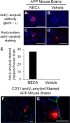
Anti-β-amyloid antibody administered intravenously crosses the BBB and labels β-amyloid plaques in transgenic mouse brains after NECA administration. A–D, Immunofluorescent microscopic images near the hippocampi of transgenic AD (APP/PSEN) mice. Mice were treated with either (A, C) NECA (0.08 mg/kg) or (B, D) vehicle and antibody to β-amyloid (6E10) was administered intravenously (top; A, B). For mice that did not receive intravenous 6E10 antibody (bottom; C, D), 6E10 was used as a primary antibody to control for the presence of plaques and was applied ex vivo during immunostaining. Blue, DAPI; red, Cy5-antibody labeling 6E10-labeled β-amyloid plaques. Scale bar, 50 μm. E, Quantification of 6E10-labeled amyloid plaques per mouse brain section in transgenic AD mice treated with NECA or vehicle alone. F, G, Immunofluorescent microscopic images of the hippocampal and cortical regions from the brains of transgenic AD mice showing an overview (F) and close-up (G) of β-amyloid plaque locations relative to blood vessels (endothelial cells are CD31 stained, green; β-amyloid plaques are 6E10 stained, red; nuclei are DAPI stained, blue). Scale bars, 50 μm.
Figure 5.
Figure 5.
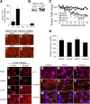
AR signaling results in changes in the paracellular but not transcellular pathway on brain endothelial cells. A, Relative genetic expression of ARs subtypes on cultured mouse BECs (Bend.3). B, Western blot analysis of A1 (left) and A2A (right) AR expression in cultured mouse BECs (Bend.3). C, AR activation decreases TEER in mouse BEC monolayers. Decreased transendothelial electrical resistance was observed after addition of NECA (1 μm) or Lexiscan (1 μm) treatment. Significant differences (Student's t test) from vehicle for Lexiscan, #p ≤ 0.05 and NECA, *p ≤ 0.05. Data are mean ± SEM. D–H, Bend.3 cells were incubated with fluorescently labeled albumin and either media alone (D), vehicle (E), NECA (1 μm) (F), or Lexiscan (1 μm) (G) for 30 min. Albumin uptake was visualized by fluorescence microscopy (albumin, red; DAPI-stained nuclei, blue). Scale bar, 50 μm. H, Albumin uptake is displayed as relative values compared with the media-alone control (set to 100%). Data are mean ± SEM (n = 5 fields per group). I–P, Actinomyosin stress fiber formation correlates with AR activation in cultured BECs. Phalloidin staining of Bend.3 cells reveals increased actinomyosin stress fiber formation following treatment with CCPA (1 μm) (M, N) or Lexiscan (1 μm) (O, P) when compared with media (I, J) or vehicle (K, L) alone. Left, 3 min treatment; right, 30 min treatment. Scale bar, 50 μm. Q–Y, AR activation induces changes in tight junction adhesion molecules in cultured BECs. ZO-1 (Q–S), Claudin-5 (T–V), and Occludin (W–Y) staining of Bend.3 cells following 1 h treatment with DMSO (left column), NECA (1 μm, middle column), and Lexiscan (1 μm, right column). Adhesion molecules are pink/red; DAPI-stained nuclei are blue. Arrowheads indicate examples of discrete changes in expression. Scale bar, 20 μm.
Figure 6.
Figure 6.
Model of AR signaling and modulation of BBB permeability. i, Basal conditions favor a tight barrier. ii, Activation of the A1 or A2A AR results in increased BBB permeability. iii, Activation of both A1 and A2A ARs results in even more permeability than observed after activation of either receptor alone.

Comment in

References

    1. Abbott NJ, Patabendige AA, Dolman DE, Yusof SR, Begley DJ. Structure and function of the blood-brain barrier. Neurobiol Dis. 2010;37:13–25. - PubMed
    1. Bartus RT, Elliott P, Hayward N, Dean R, McEwen EL, Fisher SK. Permeability of the blood brain barrier by the bradykinin agonist, RMP-7: evidence for a sensitive, auto-regulated, receptor-mediated system. Immunopharmacology. 1996;33:270–278. - PubMed
    1. Bidros DS, Vogelbaum MA. Novel drug delivery strategies in neuro-oncology. Neurotherapeutics. 2009;6:539–546. - PMC - PubMed
    1. Borlongan CV, Emerich DF. Facilitation of drug entry into the CNS via transient permeation of blood brain barrier: laboratory and preliminary clinical evidence from bradykinin receptor agonist, Cereport. Brain Res Bull. 2003;60:297–306. - PubMed
    1. Bours MJ, Swennen EL, Di Virgilio F, Cronstein BN, Dagnelie PC. Adenosine 5′-triphosphate and adenosine as endogenous signaling molecules in immunity and inflammation. Pharmacol Ther. 2006;112:358–404. - PubMed

Publication types

MeSH terms