Characterization of wild-type and alternate transcription termination signals in the Rift Valley fever virus genome
- PMID: 21917943
- PMCID: PMC3209356
- DOI: 10.1128/JVI.05322-11
Characterization of wild-type and alternate transcription termination signals in the Rift Valley fever virus genome
Abstract
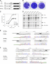
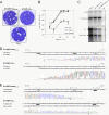
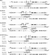
Rift Valley fever (RVF) is a mosquito-borne zoonotic disease caused by a phlebovirus of the family Bunyaviridae, which affects humans and ruminants in Africa and the Middle East. RFV virus (RVFV) possesses a single-stranded tripartite RNA genome of negative/ambisense polarity. The S segment utilizes the ambisense strategy and codes for two proteins, the N nucleoprotein and the nonstructural NSs protein, in opposite orientations. The two open reading frames (ORFs) are separated by an intergenic region (IGR) highly conserved among strains and containing a motif, 5'-GCUGC-3', present on the genome and antigenome, which was shown previously to play a role in transcription termination (C. G. Albarino, B. H. Bird, and S. T. Nichol, J. Virol. 81:5246-5256, 2007; T. Ikegami, S. Won, C. J. Peters, and S. Makino, J. Virol. 81:8421-8438, 2007). Here, we created recombinant RVFVs with mutations or deletions in the IGR and showed that the substitution of the motif sequence by a series of five A's inactivated transcription termination at the wild-type site but allowed the transcriptase to recognize another site with the consensus sequence present in the opposite ORF. Similar situations were observed for mutants in which the motif was still present in the IGR but located close to the stop codon of the translated ORF, supporting a model in which transcription is coupled to translation and translocating ribosomes abrogate transcription termination. Our data also showed that the signal tolerated some sequence variations, since mutation into 5'-GCAGC-3' was functional, and 5'-GUAGC-3' is likely the signal for the termination of the 3' end of the L mRNA.
Figures


Similar articles
-
Molecular biology and genetic diversity of Rift Valley fever virus.Antiviral Res. 2012 Sep;95(3):293-310. doi: 10.1016/j.antiviral.2012.06.001. Epub 2012 Jun 16. Antiviral Res. 2012. PMID: 22710362 Free PMC article. Review.
-
A shared transcription termination signal on negative and ambisense RNA genome segments of Rift Valley fever, sandfly fever Sicilian, and Toscana viruses.J Virol. 2007 May;81(10):5246-56. doi: 10.1128/JVI.02778-06. Epub 2007 Feb 28. J Virol. 2007. PMID: 17329326 Free PMC article.
-
Nucleocapsids of the Rift Valley fever virus ambisense S segment contain an exposed RNA element in the center that overlaps with the intergenic region.Nat Commun. 2024 Sep 1;15(1):7602. doi: 10.1038/s41467-024-52058-2. Nat Commun. 2024. PMID: 39217162 Free PMC article.
-
Characterization of Rift Valley fever virus transcriptional terminations.J Virol. 2007 Aug;81(16):8421-38. doi: 10.1128/JVI.02641-06. Epub 2007 May 30. J Virol. 2007. PMID: 17537856 Free PMC article.
-
[Rift Valley fever virus].Uirusu. 2004 Dec;54(2):229-35. doi: 10.2222/jsv.54.229. Uirusu. 2004. PMID: 15745161 Review. Japanese.
Cited by
-
Molecular biology and genetic diversity of Rift Valley fever virus.Antiviral Res. 2012 Sep;95(3):293-310. doi: 10.1016/j.antiviral.2012.06.001. Epub 2012 Jun 16. Antiviral Res. 2012. PMID: 22710362 Free PMC article. Review.
-
Reverse Genetics System for Rift Valley Fever Virus.Methods Mol Biol. 2024;2733:101-113. doi: 10.1007/978-1-0716-3533-9_7. Methods Mol Biol. 2024. PMID: 38064029
-
Roles of the coding and noncoding regions of rift valley Fever virus RNA genome segments in viral RNA packaging.J Virol. 2012 Apr;86(7):4034-9. doi: 10.1128/JVI.06700-11. Epub 2012 Jan 25. J Virol. 2012. PMID: 22278239 Free PMC article.
-
A conserved virus-induced cytoplasmic TRAMP-like complex recruits the exosome to target viral RNA for degradation.Genes Dev. 2016 Jul 15;30(14):1658-70. doi: 10.1101/gad.284604.116. Genes Dev. 2016. PMID: 27474443 Free PMC article.
-
The small genome segment of Bunyamwera orthobunyavirus harbours a single transcription-termination signal.J Gen Virol. 2012 Jul;93(Pt 7):1449-1455. doi: 10.1099/vir.0.042390-0. Epub 2012 Apr 18. J Gen Virol. 2012. PMID: 22513389 Free PMC article.
References
-
- Barr J. N., Rodgers J. W., Wertz G. W. 2006. Identification of the Bunyamwera bunyavirus transcription termination signal. J. Gen. Virol. 87:189–198 - PubMed
Publication types
MeSH terms
Substances
Grants and funding
LinkOut - more resources
Full Text Sources

