. 2011 Dec;85(23):12721-32.
doi: 10.1128/JVI.00349-11. Epub 2011 Sep 14.

Novel pentameric structure of the diarrhea-inducing region of the rotavirus enterotoxigenic protein NSP4

Affiliations

Novel pentameric structure of the diarrhea-inducing region of the rotavirus enterotoxigenic protein NSP4

Anita R Chacko et al. J Virol. 2011 Dec.

Abstract

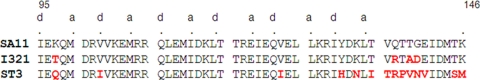
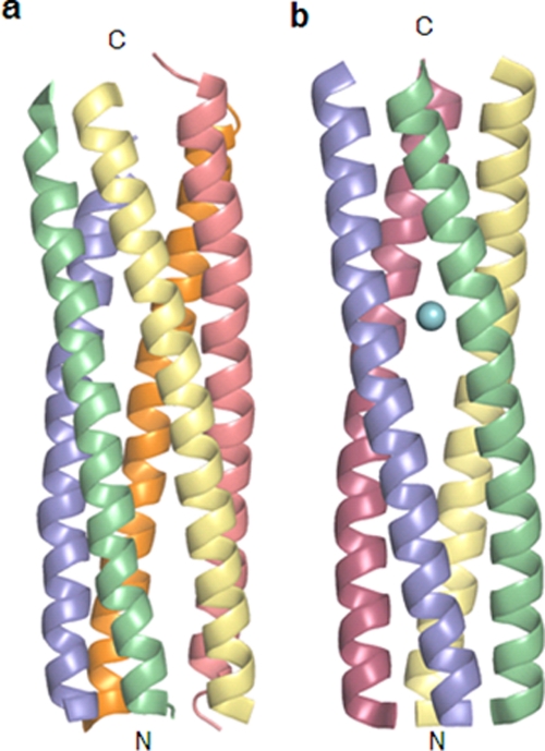
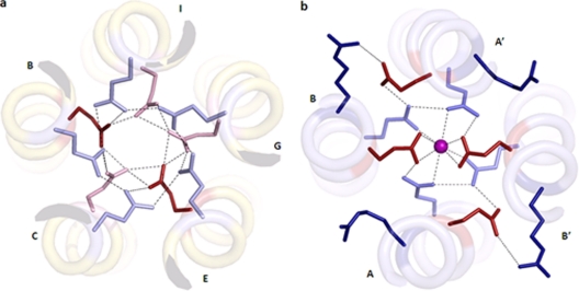
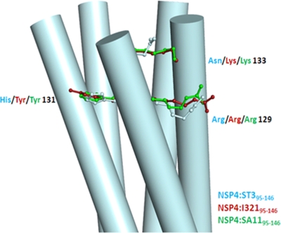
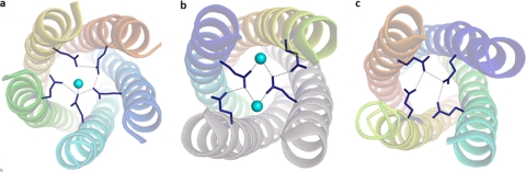
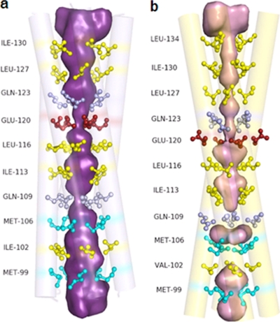
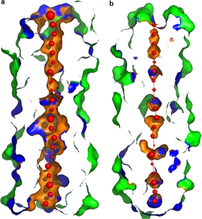
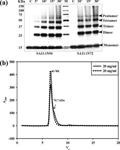
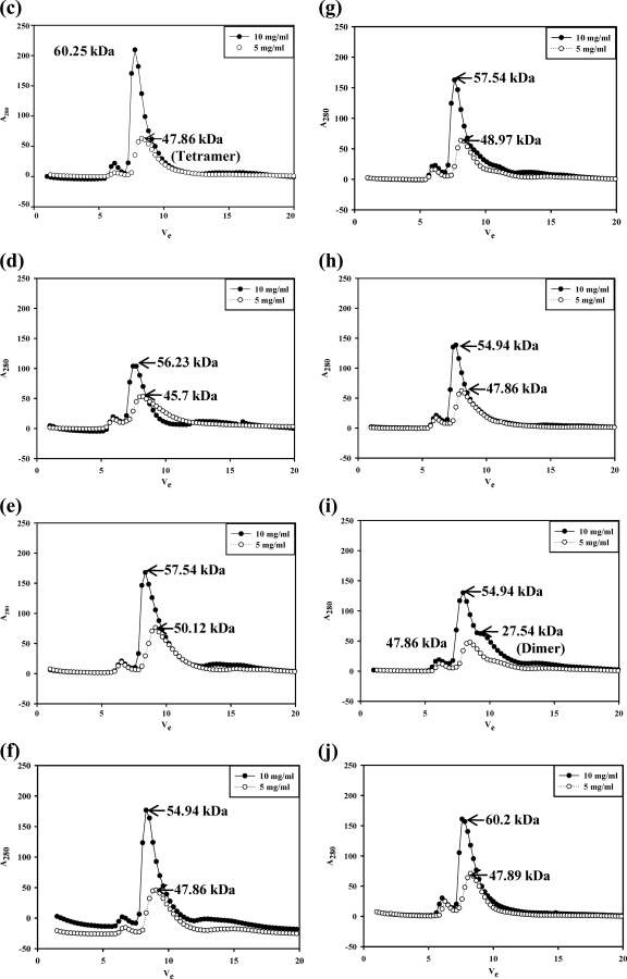
A novel pentameric structure which differs from the previously reported tetrameric form of the diarrhea-inducing region of the rotavirus enterotoxin NSP4 is reported here. A significant feature of this pentameric form is the absence of the calcium ion located in the core region of the tetrameric structures. The lysis of cells, the crystallization of the region spanning residues 95 to 146 of NSP4 (NSP4(95-146)) of strain ST3 (ST3:NSP4(95-146)) at acidic pH, and comparative studies of the recombinant purified peptide under different conditions by size-exclusion chromatography (SEC) and of the crystal structures suggested pH-, Ca(2+)-, and protein concentration-dependent oligomeric transitions in the peptide. Since the NSP4(95-146) mutant lacks the N-terminal amphipathic domain (AD) and most of the C-terminal flexible region (FR), to demonstrate that the pentameric transition is not a consequence of the lack of the N- and C-terminal regions, glutaraldehyde cross-linking of the ΔN72 and ΔN94 mutant proteins, which contain or lack the AD, respectively, but possess the complete C-terminal FR, was carried out. The results indicate the presence of pentamers in preparations of these longer mutants. Detailed SEC analyses of ΔN94 prepared under different conditions, however, revealed protein concentration-dependent but metal ion- and pH-independent pentamer accumulation at high concentrations which dissociated into tetramers and lower oligomers at low protein concentrations. While calcium appeared to stabilize the tetramer, magnesium in particular stabilized the dimer. ΔN72 existed primarily in the multimeric form under all conditions. These findings of a calcium-free NSP4 pentamer and its concentration-dependent and largely calcium-independent oligomeric transitions open up a new dimension in an understanding of the structural basis of its multitude of functions.


Figures

Fig. 1.
Fig. 1.
Sequence alignment of residues 95 to 146 of NSP4 peptides from different rotaviral strains whose crystal structures are available, showing the a and d positions of the heptad. Highlighted in red are the residues which differ from the sequence of SA11:NSP495–146. GenBank accession numbers for the NSP4 peptides of strains SA11, I321, and ST3 are JF791804, AF165066, and U59110, respectively. Note the C129R and M135T amino acid substitutions in the I321 clone used for protein expression compared to the previously reported sequence (25).
Fig. 2.
Fig. 2.
Two oligomeric structures of the coiled-coil domain of NSP4. Shown are the pentameric structure of ST3:NSP495–146 presented in this paper (a) and the tetrameric structure of SA11:NSP495–146 reported previously (PDB accession number 2O1K) (b). The bound calcium ion is shown as a sphere.
Fig. 3.
Fig. 3.
(a) Two polar layers at the helical core of the ST3:NSP495–146 pentamer. Interactions between Glu120 (red, normal pKa; pink, high pKa) and Gln123 (light blue) are marked as dotted lines. (b) SA11:NSP495–137 tetramer. The calcium ion is shown as a sphere. The ionic interactions between Arg119 (dark blue) and Glu120 (red) and hydrogen bonds between Glu120 (red) and Gln123 (light blue) and the calcium ion are marked as dotted lines.
Fig. 4.
Fig. 4.
The C-terminal end of the coiled-coil domain of the pentameric structure of NSP4:ST395–146. Residues at positions 129, 131, and 133 of one of the chains of the pentamer are shown, and the same residues from two tetrameric structures are superposed on these residues.
Fig. 5.
Fig. 5.
Interactions in the Gln109 polar layer. The water molecules are shown as cyan spheres. Interactions between Gln109 (blue) and the water molecule are marked as dotted lines. (a) ST3:NSP495–146 (pentamer); (b) SA11:NSP495–146 (dimer of dimers); (c) I321:NSP495–146 (tetramer).
Fig. 6.
Fig. 6.
The pore in the ST3:NSP495–146 pentamer (a) and the SA11:NSP495–146 tetramer (b). Shown in a ball-and-stick representation are the residues at the a and d positions of the heptad, as indicated in Fig. 1.
Fig. 7.
Fig. 7.
Surface representation of the pore in the pentameric (a) and tetrameric (b) structures of NSP495–146. Red spheres represent pore centers at 3-Å steps, with their diameters proportional to the measured diameters.
Fig. 8.
Fig. 8.
Analytical size-exclusion chromatogram of ST3:NSP495–146 under different conditions. (a) Lysis of cells in NaAc buffer (pH 5.6) (condition A). (b) Lysis in the absence of calcium (condition B). (c) Lysis in the presence of 10 mM calcium (condition C). (d) Lysis and purification in the presence of 0.1 mM calcium (condition D). (e) Lysis and purification in the presence of 0.1 mM magnesium (condition F). (f) SEC of the peptide purified under condition B at concentrations of 20 mg/ml and 5 mg/ml and under condition D at a concentration of 5 mg/ml. The NSP4 peptide in the bacterial cell lysate was precipitated in 40% ammonium sulfate. All subsequent purification steps, irrespective of cell lysis conditions, were carried out under physiological buffer conditions in the absence or presence of added calcium or magnesium. The protein was purified by ion-exchange chromatography followed by size-exclusion chromatography, and the molecular masses were determined as described in Materials and Methods. Vo, void volume.
Fig. 9.
Fig. 9.
NSP4 exists in multiple oligomeric states, as demonstrated by glutaraldehyde cross-linking and analytical gel filtration of the SA11ΔN72 and SA11ΔN94 proteins by use of a Superdex G75 column. (a) Glutaraldehyde cross-linking. Bacterial cells expressing ΔN94 were lysed in a buffer containing 10 mM Tris-HCl (pH 7.5) and 100 mM NaCl in the absence of calcium, and bacterial cells expressing ΔN72 were lysed in the presence of 10 mM CaCl2, but both were purified further under identical conditions in the absence of added calcium. The proteins were cross-linked for the indicated time periods directly on the beads using an equimolar ratio of protein to cross-linker. The proteins were resolved by SDS-PAGE, and the bands were visualized by silver staining. Note the pentamers and other oligomeric forms of both mutant proteins purified after the lysis of cells in the presence or absence of calcium. M, Precision Plus protein standards from Bio-Rad. (b) Analytical size-exclusion chromatography of ΔN72. The protein was analyzed after purification from cells lysed in Tris-HCl (pH 7.5) buffer in the presence (−) or absence (.....) of 10 mM CaCl2. Vo, void volume. (c to j) Analytical size-exclusion chromatography of ΔN94 purified under different conditions. (c) Cells lysed in NaAc buffer (pH 5.6) (condition A); (d) lysis and purification in the absence of calcium (condition B); (e) lysis in the presence of 0.1 mM CaCl2 (condition C0.1); (f) lysis in the presence of 1.0 mM CaCl2 (condition C1); (g) lysis in the presence of 10 mM CaCl2 (condition C10); (h) lysis and purification in the presence of 0.1 mM CaCl2 (condition D); (i) lysis in the presence of 10 mM MgCl2 (condition E); (j) lysis and purification in the presence of 0.1 mM MgCl2 (condition F).
Fig. 9.
Fig. 9.
NSP4 exists in multiple oligomeric states, as demonstrated by glutaraldehyde cross-linking and analytical gel filtration of the SA11ΔN72 and SA11ΔN94 proteins by use of a Superdex G75 column. (a) Glutaraldehyde cross-linking. Bacterial cells expressing ΔN94 were lysed in a buffer containing 10 mM Tris-HCl (pH 7.5) and 100 mM NaCl in the absence of calcium, and bacterial cells expressing ΔN72 were lysed in the presence of 10 mM CaCl2, but both were purified further under identical conditions in the absence of added calcium. The proteins were cross-linked for the indicated time periods directly on the beads using an equimolar ratio of protein to cross-linker. The proteins were resolved by SDS-PAGE, and the bands were visualized by silver staining. Note the pentamers and other oligomeric forms of both mutant proteins purified after the lysis of cells in the presence or absence of calcium. M, Precision Plus protein standards from Bio-Rad. (b) Analytical size-exclusion chromatography of ΔN72. The protein was analyzed after purification from cells lysed in Tris-HCl (pH 7.5) buffer in the presence (−) or absence (.....) of 10 mM CaCl2. Vo, void volume. (c to j) Analytical size-exclusion chromatography of ΔN94 purified under different conditions. (c) Cells lysed in NaAc buffer (pH 5.6) (condition A); (d) lysis and purification in the absence of calcium (condition B); (e) lysis in the presence of 0.1 mM CaCl2 (condition C0.1); (f) lysis in the presence of 1.0 mM CaCl2 (condition C1); (g) lysis in the presence of 10 mM CaCl2 (condition C10); (h) lysis and purification in the presence of 0.1 mM CaCl2 (condition D); (i) lysis in the presence of 10 mM MgCl2 (condition E); (j) lysis and purification in the presence of 0.1 mM MgCl2 (condition F).

Similar articles

Cited by

References

    1. Adams P. D., et al. 2002. Crystallographic structure determination. Acta Crystallogr. D 58:1948–1954 - PubMed
    1. Au K. S., Chan W. K., Burns J. W., Estes M. K. 1989. Receptor activity of rotavirus nonstructural glycoprotein NS 28. J. Virol. 63:4553–4562 - PMC - PubMed
    1. Ball J. M., Tian P., Zeng C. Q.-Y., Morris A. P., Estes M. K. 1996. Age-dependent diarrhea induced by a rotaviral nonstructural glycoprotein. Science 272:101–104 - PubMed
    1. Bas D. C., Rogers D. M., Jensen J. H. 2008. Very fast prediction and rationalization of pKa values for protein-ligand complexes. Proteins 73:765–783 - PubMed
    1. Bergman C. C., Maass D., Poruchynsky M. S., Atkinson P. H., Bellamy A. R. 1989. Topology of the non-structural rotavirus receptor glycoprotein NS28 in the rough endoplasmic reticulum. EMBO J. 8:1695–1703 - PMC - PubMed

Publication types

MeSH terms