. 2011 Nov;85(22):11725-31.
doi: 10.1128/JVI.05680-11. Epub 2011 Sep 14.

Differential reactivity of germ line allelic variants of a broadly neutralizing HIV-1 antibody to a gp41 fusion intermediate conformation

Affiliations

Differential reactivity of germ line allelic variants of a broadly neutralizing HIV-1 antibody to a gp41 fusion intermediate conformation

S Munir Alam et al. J Virol. 2011 Nov.

Abstract

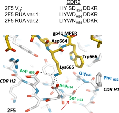
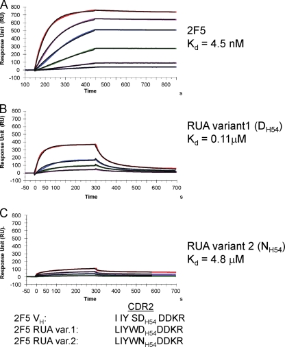
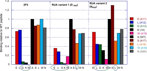
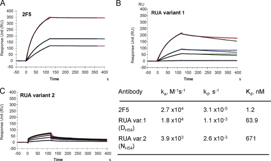
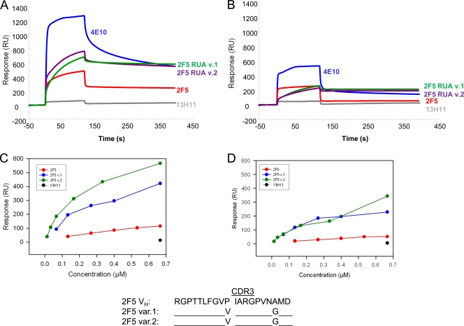
Genetic factors, as well as antigenic stimuli, can influence antibody repertoire formation. Moreover, the affinity of antigen for unmutated naïve B cell receptors determines the threshold for activation of germinal center antibody responses. The gp41 2F5 broadly neutralizing antibody (bNAb) uses the V(H)2-5 gene, which has 10 distinct alleles that use either a heavy-chain complementarity-determining region 2 (HCDR2) aspartic acid (D(H54)) or an HCDR2 asparagine (N(H54)) residue. The 2F5 HCDR2 D(H54) residue has been shown to form a salt bridge with gp41 (665)K; the V(H)2-5 germ line allele variant containing N(H54) cannot do so and thus should bind less avidly to gp41. Thus, the induction of 2F5 bNAb is dependent on both genetic and structural factors that could affect antigen affinity of unmutated naïve B cell receptors. Here, we studied allelic variants of the V(H)2-5 inferred germ line forms of the HIV-1 gp41 bNAb 2F5 for their antigen binding affinities to gp41 linear peptide and conformational protein antigens. Both V(H)2-5 2F5 inferred germ line variants bound to gp41 peptides and protein, including the fusion intermediate protein mimic, although more weakly than the mature 2F5 antibody. As predicted, the affinity of the N(H54) variant for fusion-intermediate conformation was an order of magnitude lower than that of the D(H54) V(H)2-5 germ line antibody, demonstrating that allelic variants of 2F5 germ line antibodies differentially bind to gp41. Thus, these data demonstrate a genetically determined trait that may affect host responses to HIV-1 envelope epitopes recognized by broadly neutralizing antibodies and has implications for unmutated ancestor-based immunogen design.


Figures

Fig. 1.
Fig. 1.
Bonding between 2F5 HCDR2 DH54 and gp41 MPER 665K (27). The contact surface of 2F5 bound to its antigenic peptide shows a strong complementarity of charge, and two CDR H2 residues, DH56 and DH54, interact through hydrogen bonds and salt bridges with K665 of the 2F5 core tripeptide DKW. Based on the CDR H2 substitutions in the two UAs, the following predictions can be made. First, the CDR H2 DH54 residue in variant 1 is retained and, if positioned appropriately, could form a salt bridge with K665 of the 2F5 core DKW. However, the bulky side chain of WH53 (from SH53 to WH53, red arrow) is very likely to perturb the local environment of HCDR2, through potential steric clashes with HCDR1 backbone and FH32 side chain, and disruption of the H bond with GH33. Second, the additional alteration of DH54 to NH54 in UA variant 2 HCDR2 will disrupt the salt bridge with 665K. Also, there is a potential for HCDR2 NH54 to H bond with DH56, which would further preclude establishing bonding with gp41 K665. Critical CDR residues are labeled in green, HCDR1 residues are labeled in blue, and gp41 MPER residues are labeled in black. gp41 MPER is shown in yellow, and 2F5 antibody backbone is shown in white.
Fig. 2.
Fig. 2.
2F5 UAs bind to 2F5 nominal epitope peptide with weaker affinity than 2F5 MAb. The gp41 MPER peptide (QQEKNEQELLELDKWASLWN), which includes the 2F5 nominal epitope peptide, was anchored (200 to 300 RU) on a streptavidin (SA) chip via a biotin tag attached to the N terminus of the peptide. Each MAb was injected at various concentrations: 0.67, 1.7, 3.3, 6.7, 13.3, and 26.7 nM (2F5) (A); 3.3, 6.7, 13.3, and 33.3 nM (2F5 UA variant 1) (B); and 66.7, 165.7, 333.3, and 500 nM (2F5 UA variant 2) (C). UA variant 1 with HCDR2 DH54 bound to gp41 MPER peptide with a 40-fold-higher binding Kd than that of UA variant 2 (NH54). Rate constants, ka and kd, for 2F5 were 1.97 × 10⁵ M−1 s−1 and 8.8 × 10⁻⁴ s−1; those for UA variant 1 were 3.0 × 10⁵ M−1 s−1 and 3.26 × 10⁻² s−1; those for UA variant 2 were 8.4 × 10³ M−1 s−1 and 4.1 × 10⁻² s−1, respectively. Data are representative of at least two independent experiments.
Fig. 3.
Fig. 3.
2F5 UAs show broader specificity than does the mature 2F5 antibody. The effect of alanine substitution of each of the indicated residues of the 2F5 nominal epitope peptide on the binding of 2F5, UA variant 1, or UA variant 2 is plotted as a ratio of the binding of mutant to wild-type (WT) peptide. Critical residues showing >50% reduction in steady binding responses as measured by SPR analysis are circled.
Fig. 4.
Fig. 4.
Binding of 2F5 and 2F5 UAs to trimeric gp41-inter protein. Each of the antibodies, 2F5 (A), UA variant 1 (B), or UA variant 2 (C), was captured to about 600 to 1,000 RU on individual flow cells immobilized with anti-human Fc antibody. 92UG gp41-inter protein (13) was injected over each of the captured antibodies at concentrations of 38.5, 76.9, 192.3, 384.6, and 769 nM (UA variant 1) or 769, 1,538.5, 2,307, 3,077, and 3,846 nM (UA variant 2). The rate constants and Kd values shown in the table were derived from a global curve analysis using the 1:1 Langmuir equation and are representative of two experiments.
Fig. 5.
Fig. 5.
2F5 UAs bind to anionic phospholipids. (A and B) Synthetic liposomes with either cardiolipin (CL) (A) or phosphatidylserine (PS) (B) were prepared at a percent molar ratio of 25:75 with phosphatidylcholine (PC) (PC/CL or PC/PS ratio) as described earlier (1, 2, 11). Each of the liposomes was captured to about 500 RU on an L1 sensor chip, and the indicated antibodies at 100 μg/ml were injected over the lipid captured surfaces. No binding was observed on a control PC liposome captured surface (not shown). The control was the nonneutralizing gp41 MPER antibody 13H11, which does not bind to phospholipids (1, 26). (C and D) Binding in resonance units (RU) of each antibody at various concentrations to cardiolipin (C)- or PS (D)-containing liposomes shows higher binding responses of both 2F5 UA variants than of 2F5 MAb. No binding was observed for the 13H11 MAb at the highest concentration of 100 μg/ml (C and D). 2F5 HCDR3 sequences show two substitutions in 2F5 UA HCDR3, V to P and A to G. The residue P100E is at the apex of the 2F5 HCDR3 loop (27).

References

    1. Alam S. M., et al. 2007. The role of antibody polyspecificity and lipid reactivity in binding of broadly neutralizing anti-HIV-1 envelope human monoclonal antibodies 2F5 and 4E10 to glycoprotein 41 membrane proximal envelope epitopes. J. Immunol. 178: 4424–4435 - PMC - PubMed
    1. Alam S. M., et al. 2009. Role of HIV membrane in neutralization by two broadly neutralizing antibodies. Proc. Natl. Acad. Sci. U. S. A. 106: 20234–20239 - PMC - PubMed
    1. Alam S. M., et al. 2008. Human immunodeficiency virus type 1 gp41 antibodies that mask membrane proximal region epitopes: antibody binding kinetics, induction, and potential for regulation in acute infection. J. Virol. 82: 115–125 - PMC - PubMed
    1. Berman J. E., Alt F. W. 1990. Human heavy chain variable region gene diversity, organization, and expression. Int. Rev. Immunol. 5: 203–214 - PubMed
    1. Cardoso R. M., et al. 2005. Broadly neutralizing anti-HIV antibody 4E10 recognizes a helical conformation of a highly conserved fusion-associated motif in gp41. Immunity 22: 163–173 - PubMed

Publication types

MeSH terms

Substances