

doi: 10.1128/JVI.05970-11.
Epub 2011 Sep 14.
Mutations in the membrane-proximal region of the influenza A virus M2 protein cytoplasmic tail have modest effects on virus replication
Affiliations
- PMID: 21917980
- PMCID: PMC3209349
- DOI: 10.1128/JVI.05970-11
Item in Clipboard
Mutations in the membrane-proximal region of the influenza A virus M2 protein cytoplasmic tail have modest effects on virus replication
J Virol.
2011 Dec.
Abstract
Influenza A virus encodes M2, a proton channel that has been shown to be important during virus entry and assembly. In order to systematically investigate the role of the membrane-proximal residues in the M2 cytoplasmic tail in virus replication, we utilized scanning and directed alanine mutagenesis in combination with transcomplementation assays and recombinant viruses. The membrane-proximal residues 46 to 69 tolerated numerous mutations, with little, if any, effect on virus replication, suggesting that protein structure rather than individual amino acid identity in this region may be critical for M2 protein function.
Figures
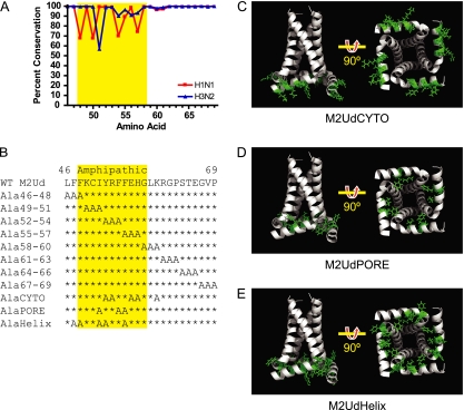
Sequence and structural location of M2 cytoplasmic tail amino acids. (A) Conservation of residues 46 to 69 of the M2 protein among H1N1 (virus sequences prior to 2009) and H3N2 influenza A virus strains. (B) Sequences of residues 46 to 69 in wild-type M2 protein and alanine substitution mutants. The amphipathic helix is highlighted in yellow (34). (C to E) Structures of M2 mutant proteins (residues 22 to 62), with mutated amino acids shown in green. Residue 22 appears at the top of the structures on the left. The structures on the right are rotated 90°, as indicated, in order to show the cytosolic face of the M2 tetramer. (C) M2UdCYTO mutant (mutations in Y52, R53, E56, H57, and K60). (D) M2UdPORE mutant (mutations in I51, F54, and F55). (E) M2UdHelix mutant (mutations in F47, F48, I51, Y52, and F55). Structures were generated based on the structure under Protein Data Bank accession number 2LOJ by using PyMol.
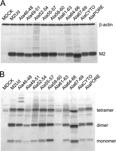
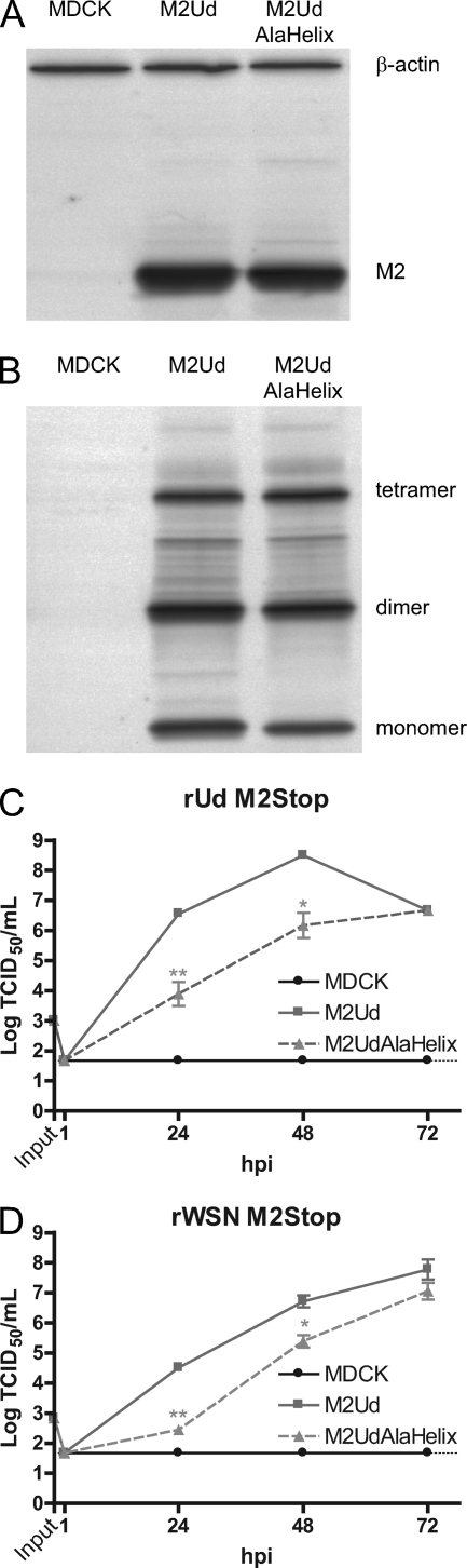
Expression and oligomerization of mutant M2 proteins. MDCK cells stably expressing the indicated M2 proteins were analyzed by Western blotting in order to determine the overall expression under reducing conditions (A) and the presence of disulfide-linked oligomers under nonreducing conditions (B). Monomeric M2 and the cell protein loading control, β-actin, are indicated in panel A, and monomers, dimers, and tetramers of M2 are indicated in panel B.
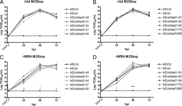
Complementation of M2-deficient viruses by expression of mutant M2 proteins. MDCK cells stably expressing the indicated M2 proteins were infected at an MOI of 0.001 with a recombinant influenza A virus that does not encode the full-length M2 protein. The amount of infectious virus at each time point was determined by TCID50 assay on cells expressing wild-type M2. Complementation of rUd M2Stop virus (A and B) or rWSN M2Stop virus (C and D) infectivity was determined with cells expressing the indicated M2 proteins. The means and standard errors of the means for triplicate samples from a representative experiment are shown. The limit of detection is marked by a horizontal dotted line.
Plaque sizes and replication kinetics of recombinant viruses expressing mutated M2 proteins. (A and C) Plaque diameters of MDCK cells infected with the indicated recombinant viruses at 3 days postinfection (dpi). The cells were fixed and stained with a Naphthol Blue Black solution, and individual plaque diameters were measured from scanned images by using ImageJ. Representative data from triplicate experiments are shown. The Student t test was performed to determine significant differences compared to wild-type virus. (B and D) MDCK cells were infected with the indicated recombinant viruses at an MOI of 0.001. At the indicated hpi, cell supernatants were collected and the numbers of infectious virus particles were determined by TCID50 assay on M2-expressing cells. The means and standard errors of the means for triplicate samples from a representative experiment are shown. The limit of detection is marked by a horizontal dotted line.
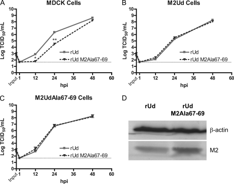
Complementation of a recombinant virus expressing M2Ala67-79. MDCK (A), M2Ud-expressing (B), and M2UdAla67-69-expressing (C) cells were infected with the indicated recombinant viruses at an MOI of 0.001. At the indicated hpi, cell supernatants were collected and the numbers of infectious virus particles were determined by TCID50 assay on cells expressing wild-type M2. The means and standard errors of the means for triplicate samples from a representative experiment are shown. The limit of detection is marked by a horizontal dotted line. (D) MDCK cells were infected with the indicated recombinant viruses at an MOI of 0.5. At 16 hpi, expression in cell lysates of M2 and the cell protein loading control, β-actin, was compared by Western blotting.
Expression and function of M2UdAlaHelix protein. Western blot analysis was performed with MDCK cells stably expressing either wild-type M2Ud or M2UdAlaHelix. (A) Total expression of M2 protein under reducing conditions. (B) Presence of disulfide-linked oligomers under nonreducing conditions. Monomeric M2 and the cell protein loading control, β-actin, are indicated in panel A, and monomers, dimers, and tetramers of M2 are indicated in panel B. (C and D) MDCK cells stably expressing the indicated M2 protein were infected with the indicated recombinant virus that does not encode the full-length M2 protein at an MOI of 0.001. The amount of infectious virus at each time point was determined by TCID50 assay on cells expressing wild-type M2. The means and standard errors of the means for triplicate samples from a representative experiment are shown. The limit of detection is marked by a horizontal dotted line.
Characterization of a recombinant virus expressing M2AlaHelix. (A) Plaque diameters of MDCK cells infected with recombinant viruses expressing either wild-type M2 or M2AlaHelix at 3 dpi. The individual and average diameters of plaques were determined. Representative data from triplicate experiments are shown. (B) MDCK cells were infected with the indicated viruses at an MOI of 0.001. At the indicated hpi, cell supernatants were collected and the numbers of infectious virus particles were determined by TCID50 assay on M2-expressing cells. The means and standard errors of the means for triplicate samples from a representative experiment are shown. The limit of detection is marked by a horizontal dotted line. (C) MDCK cells infected with 500,000 TCID50 of the indicated virus were stained for immunofluorescence at 15 hpi and visualized by microscopy. The percentage and average percentage of infected cells showing filaments were determined from 20 nonoverlapping images taken with an epifluorescence microscope. Data for one representative experiment are shown. (D) MDCK cells were infected with the indicated virus at an MOI of 5. At 12 hpi, supernatants were collected, virus particles were concentrated by ultracentrifugation through 35% sucrose in PBS and resuspended in PBS, and equal volumes were analyzed along with the cell lysates by Western blotting under reducing conditions.
References
-
- Bourmakina S. V., Garcia-Sastre A. 2003. Reverse genetics studies on the filamentous morphology of influenza A virus. J. Gen. Virol. 84:517–527 - PubMed
Publication types
MeSH terms
Substances
Grants and funding
LinkOut - more resources
Full Text Sources
