. 2011 Dec;85(23):12673-85.
doi: 10.1128/JVI.05296-11. Epub 2011 Sep 14.

Human T-cell lymphotropic virus type 3 (HTLV-3)- and HTLV-4-derived antisense transcripts encode proteins with similar Tax-inhibiting functions but distinct subcellular localization

Affiliations

Human T-cell lymphotropic virus type 3 (HTLV-3)- and HTLV-4-derived antisense transcripts encode proteins with similar Tax-inhibiting functions but distinct subcellular localization

Émilie Larocque et al. J Virol. 2011 Dec.

Abstract

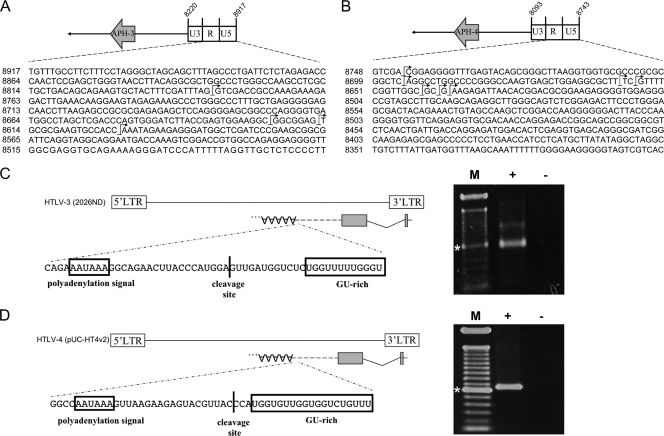
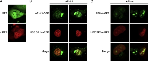
The human T-cell lymphotropic virus (HTLV) retrovirus family is composed of the well-known HTLV type 1 (HTLV-1) and HTLV-2 and the most recently discovered HTLV-3 and HTLV-4. Like other retroviruses, HTLV-1 and HTLV-2 gene expression has been thought to be orchestrated through a single transcript. However, recent reports have demonstrated the unique potential of both HTLV-1 and HTLV-2 to produce an antisense transcript. Furthermore, these unexpected and newly identified transcripts lead to the synthesis of viral proteins termed HBZ (HTLV-1 basic leucine zipper) and APH-2 (antisense protein of HTLV-2), respectively. As potential open reading frames are present on the antisense strand of HTLV-3 and HTLV-4, we tested whether in vitro antisense transcription occurred in these viruses and whether these transcripts had a coding potential. Using HTLV-3 and HTLV-4 proviral DNA constructs, antisense transcripts were detected by reverse transcriptase PCR. These transcripts are spliced and polyadenylated and initiate at multiple sites from the 3' long terminal repeat (LTR). The resulting proteins, termed APH-3 and APH-4, are devoid of a typical basic leucine zipper domain but contain basic amino acid-rich regions. Confocal microscopy and Western blotting experiments demonstrated a nucleus-restricted pattern for APH-4, while APH-3 was localized both in the cytoplasm and in the nucleus. Both proteins showed partial colocalization with nucleoli and HBZ-associated structures. Finally, both proteins inhibited Tax1- and Tax3-mediated HTLV-1 and HTLV-3 LTR activation. These results further demonstrate that retroviral antisense transcription is not exclusive to HTLV-1 and HTLV-2 and that APH-3 and APH-4 could impact HTLV-3 and HTLV-4 replication.


Figures

Fig. 1.
Fig. 1.
Position of antisense ORF in HTLV-3 and HTLV-4 genomes. The HTLV-3 2026ND (A) and HTLV-4 HT4v2 (B) molecular clones and known genes are depicted. The positions of the putative APH-3 and APH-4 ORFs from the antisense strand are indicated below each proviral DNA. Versions of these vectors with 5′ deletions, termed pHTLV-3 ΔEcoRV and pHTLV-4 ΔSacI, are also presented. The size of the proviral DNA (full-length version or version with 5′-end deletion) is indicated for each retrovirus.
Fig. 2.
Fig. 2.
Detection of spliced HTLV-3 and HTLV-4 antisense transcripts. 293T cells were transfected with either pHTLV-3 ΔEcoRV (A) or pHTLV-4 ΔSacI (B). RNA was extracted and analyzed by RT-PCR for the presence of antisense transcripts using the primer combinations LTR-HTLV-3as1/env-HTLV-3s8 (A) or LTR-HTLV-4as2/env-HTLV-4s1 (B). Spliced transcripts for each proviral DNA are depicted, and the positions of the SD and SA sites are indicated below (nucleotidic positioning from sense strand). Lanes: −, negative control (no RT added in RT step before PCR); M, 100-bp marker (*, 600 bp); +, test lane. (C) Amino acid sequence deduced from spliced APH-3 and APH-4 RNA next to the splice junction. The amino acid sequence is shown above each nucleotide sequence. The nucleotide and amino acid sequences predicted for unspliced transcripts are provided for comparison. Asterisks, in-frame stop codons. (D) Jurkat cells were transfected with pHTLV-3 ΔEcoRV or pHTLV-4 ΔSacI, and RNA was extracted and analyzed by RT-PCR as described above using the primer combination LTR-HTLV-3as1/env-HTLV-3s3 (left) or LTR-HTLV-4as2/env-HTLV-4as5 (right).
Fig. 3.
Fig. 3.
Identification of transcription initiation sites and poly(A) addition sites for APH-3 and APH-4 transcripts. Cultured 293T cells were transfected with versions of either pHTLV-3 (nt 4731 to 8918 containing the 3′ LTR and the antisense ORF) (A and C) or pHTLV-4 (nt 4873 to 8742 containing the 3′LTR and the antisense ORF) (B and D) from which proviral DNA was deleted. RNA was extracted at 48 h posttransfection and analyzed by 5′ or 3′ RLM-RACE. (A and B) The antisense nucleotide sequence of the 3′ LTR is depicted, and initiation sites are shown by arrows. (C and D) The positions of the APH-3 and APH-4 mRNAs (grey boxes) and of the 3′ poly(A) tails are shown. In the sequence below, the position of the cleavage site and the sequences of the poly(A) signal and GU-rich elements are shown. Lanes: +, test lane; −, PCR amplification with no prior RT step; M, 100-bp marker (*, 600 bp).
Fig. 4.
Fig. 4.
Predicted amino acid sequences of APH-3 and APH-4. The amino acid sequences of APH-3 and APH-4 were deduced from the resulting antisense transcripts produced by the 2026ND (HTLV-3) and HT4v2 (HTLV-4) strains, respectively. Both sequences were compared to the amino acid sequence of the HBZ-SP1 isoform and to the previously reported sequence of the HTLV-2-derived APH-2 antisense protein. The described BR2, BR1, and DNA-binding domain (DBD) are indicated above the HBZ sequence, while the LZ domain is displayed in a box. LXXLL and LXXLL-like motifs are underlined in both APH-3 and APH-4 amino acid sequences.
Fig. 5.
Fig. 5.
APH-3 and APH-4 demonstrate different cellular localizations. (A and B) pMycAPH-3 and pMycAPH-4 expression vectors were transfected into COS-7 (A) or Jurkat (B) cells. At 36 h posttransfection, cells were fixed and stained as described in Materials and Methods. Cells were mounted in ProLong antifade reagent in the presence of propidium iodide. Images are representative of those for the entire population of transfected cells. Panels on the right show the merging of both Myc (green) and PI (red) signals. Samples were observed with a Bio-Rad laser scanning confocal microscope (MRC-1024ES) with a ×60 objective under oil immersion and with a numerical aperture of 1.4. Images were acquired with the LaserSharp software. (C) The parental peGFP-N1, pAPH-3–GFP, and pAPH-4–GFP expression vectors were transfected into COS-7 cells. At 36 h posttransfection, cells were visualized by confocal microscopy. Panels on the left show the GFP signal, the middle panels show phase-contrast, and the panels on the right show merging of both the phase-contrast and GFP signals. (D) pAPH-3–GFP and pAPH-4–GFP expression vectors were cotransfected into COS-7 cells with pNucleolin-dsRed. At 48 h posttransfection, cells were observed by confocal microscopy. Panels on the left show the GFP signal, the middle panels show the Nucleolin-dsRed signal, and the panels on the right show merged GFP and Nucleolin-dsRed signals. Samples were maintained in supplemented DMEM during observation. For both panels C and D, a Bio-Rad laser scanning confocal microscope (MRC-1024ES) was used to visualize positive cells with a ×20 objective under water immersion and with a numerical aperture of 0.75. (E) Cell lysates (50 μg) from 293T cells transfected with pHBZ-Myc, pMycAPH-3, or pMycAPH-4 were separated into cytoplasmic (Cyt) and nuclear (Nuc) fractions, subjected to electrophoresis on a 12% bis-Tris gel, and analyzed by Western blotting with anti-Myc, anti-α-tubulin, and anti-Lap-2 antibodies. Cells transfected with the pcDNA3.1/Myc parental vector served as a negative control (pCDNA3.1). (F) Cell lysates (50 μg) from COS-7 cells transfected with pHBZ-Myc, pMycAPH-3, or pMycAPH-4 were separated into cytoplasmic (Cyt) and nuclear (Nuc) fractions, subjected to electrophoresis on a 12% bis-Tris gel, and analyzed by Western blotting with anti-Myc and anti-α-tubulin antibodies.
Fig. 6.
Fig. 6.
Partial colocalization of APH-3 and APH-4 with HBZ. (A) Parental vectors peGFP-N1 and pcDNA3.1Zeo(+)-mRFP were transfected into COS-7 cells and visualized by confocal microscopy. (B and C) pAPH-3–GFP (B) and pAPH-4–GFP (C) expression vectors were cotransfected with HBZ SP1-mRFP into COS-7 cells. At 48 h posttransfection, cells were observed by confocal microscopy. Panels on the top show the GFP signal, middle panels show the mRFP signal, and panels at the bottom show merging of both GFP and mRFP signals. Samples were maintained in supplemented DMEM during observation. A Bio-Rad laser scanning confocal microscope (MRC-1024ES) was used to visualize positive cells with a ×20 objective under water immersion and with a numerical aperture of 0.75.
Fig. 7.
Fig. 7.
Antisense expression is upregulated in Jurkat T cells after activation. (A) The firefly luciferase reporter gene was inserted in frame with exon 2 of both APH-3 and APH-4 along with a poly(A) signal cassette at the 3′ end to generate pHTLV-3-as-luc and pHTLV-4-as-luc. (B) Both luciferase-encoding constructs were transfected into Jurkat T cells and were then stimulated for 8 h with PHA, PMA, ionomycin, bpV[pic], forskolin, TNF-α, or different combinations of these agents. Luciferase activity was measured from cell lysates of three independently stimulated samples, and the average fold induction (± standard deviation) is presented, where unstimulated cells are assigned a value of 1. These results are representative of four independent experiments.
Fig. 8.
Fig. 8.
APH-3 and APH-4 repress Tax1- and Tax3-dependent transactivation of the HTLV-1 LTR. (A) 293T cells were transiently cotransfected with pHTLV-1 luc and pCMVTax1 together with 0.2 μg or 0.4 μg pHBZ-SP1-Myc, pMycAPH-3, pMycAPH-4, or the empty vector pcDNA3.1 and pRcActin-lacZ. Cells were lysed at 48 h posttransfection. These results represent those from three independent experiments. (Bottom set of panels) Cell lysates (25 μg) were prepared from these transfections and analyzed by Western blotting using mouse anti-Myc (upper), anti-Tax serum (middle), and mouse anti-GAPDH (bottom). (B) 293T cells were transiently transfected with pHTLV-1 luc and pCMVTax3 together with pHBZ-SP1-Myc, pMycAPH-3, pMycAPH-4, or the empty vector pcDNA3.1 and pRcActin-lacZ. (C) 293T cells were transiently cotransfected with pHTLV-3 luc and pCMVTax3 along with pHBZ-SP1-Myc, pMycAPH-3, pMycAPH-4, or the empty vector pcDNA3.1 and pRcActin-lacZ. The presented luciferase activities are averages of three independent transfection experiments and are depicted as the average normalized luciferase activity ± standard deviation. These results represent those from three independent experiments.

References

    1. Arnold J., et al. 2006. Enhancement of infectivity and persistence in vivo by HBZ, a natural antisense coded protein of HTLV-1. Blood 107:3976–3982 - PMC - PubMed
    1. Arnold J., Zimmerman B., Li M., Lairmore M. D., Green P. L. 2008. Human T-cell leukemia virus type-1 antisense-encoded gene, Hbz, promotes T lymphocyte proliferation. Blood 112:3788–3797 - PMC - PubMed
    1. Arpin-Andre C., Mesnard J. M. 2007. The PDZ domain-binding motif of the human T cell leukemia virus type 1 tax protein induces mislocalization of the tumor suppressor hScrib in T cells. J. Biol. Chem. 282:33132–33141 - PubMed
    1. Barbeau B., Mesnard J. M. 2007. Does the HBZ gene represent a new potential target for the treatment of adult T-cell leukemia? Int. Rev. Immunol. 26:283–304 - PubMed
    1. Barbeau B., Robichaud G. A., Fortin J.-F., Tremblay M. J. 2001. Negative regulation of the NFAT1 factor by CD45: implication in HIV-1 LTR activation. J. Immunol. 167:2700–2713 - PubMed

Publication types

MeSH terms