Review
. 2011 Dec 1;3(12):a005058.
doi: 10.1101/cshperspect.a005058.

Extracellular matrix degradation and remodeling in development and disease

Affiliations
Review

Extracellular matrix degradation and remodeling in development and disease

Pengfei Lu et al. Cold Spring Harb Perspect Biol. .

Abstract

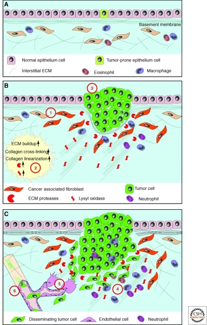
The extracellular matrix (ECM) serves diverse functions and is a major component of the cellular microenvironment. The ECM is a highly dynamic structure, constantly undergoing a remodeling process where ECM components are deposited, degraded, or otherwise modified. ECM dynamics are indispensible during restructuring of tissue architecture. ECM remodeling is an important mechanism whereby cell differentiation can be regulated, including processes such as the establishment and maintenance of stem cell niches, branching morphogenesis, angiogenesis, bone remodeling, and wound repair. In contrast, abnormal ECM dynamics lead to deregulated cell proliferation and invasion, failure of cell death, and loss of cell differentiation, resulting in congenital defects and pathological processes including tissue fibrosis and cancer. Understanding the mechanisms of ECM remodeling and its regulation, therefore, is essential for developing new therapeutic interventions for diseases and novel strategies for tissue engineering and regenerative medicine.


Figures

Figure 1.
Figure 1.
Regulation of ECM remodeling enzymes and biological consequences of ECM dynamics. (A) Some of the mechanisms whereby activities of ECM remodeling enzymes are regulated. The spatiotemporal expression of ECM remodeling enzymes is regulated by transcription factors (1). Once expressed, enzymes may be delivered to specific subcellular locations, including the migration front of a cell. Depending on whether they carry a transmembrane domain, enzymes may anchor in the plasma membrane or be secreted (2). When initially produced, most ECM remodeling enzymes exist as precursors that are inactive until processed and the autoinhibitory prodomain is removed by other proteases (3). Active enzymes can be quickly neutralized by endogenous specific or paninhibitors (4), which then are subject to permanent removal via degradation in the lysosomes (5). (B) The versatile functions of the ECM depend on its diverse physical, biochemical, and biomechanical properties. Anchorage to the basement membrane is essential for various biological processes, including asymmetric cell division in stem cell biology and maintenance of tissue polarity (1). Depending on contexts, the ECM may serve as a barrier (2) or facilitator to cell migration (3). In addition, by binding to growth factor signaling molecules and preventing their otherwise free diffusion, the ECM acts as a sink for these signals and helps shape a concentration gradient (4). Certain ECM components, including heparan sulfate and ECM receptors such as CD44, can selectively bind to different growth factors and function as signal “coreceptors” (5) or “presenters” (6) and help determine the direction of cell–cell communication. Finally, ECM biomechanical properties, including stiffness, have profound influences on various cell behaviors, including cell differentiation (7).
Figure 2.
Figure 2.
ECM dynamics determine epithelial branch patterning in vertebrate organs. The ECM is dynamic and plays essential roles in various steps during vertebrate epithelial branching morphogenesis. Deposition of newly synthesized ECM (green solid line) including fibronectin and laminin is required for splitting the epithelial bud and primary branching (1). In contrast, partial degradation of the ECM (gray dotted line) by MMP is necessary for epithelial cells to sprout from the side of the duct and undergo side branching (2). MMP activities are also required at the invasion front to maintain a constant ECM remodeling process that is essential for collective epithelial migration (3). MMP activities also generate functional ECM fragments to promote cell proliferation in the tip epithelial cells and thus are essential for supplying the necessary building blocks to sustain the rapid progress of epithelial branching. Interestingly, newly synthesized ECM is also deposited around the “neck” of the branching tip (4). ECM deposition at this place may be important for the ductal remodeling process that has been observed in kidney epithelial branching. (Figure was revised from the original version created by Mark Sternlicht [Sternlicht et al. 2006] and reprinted, with permission, from Elsevier © 2006.)
Figure 3.
Figure 3.
ECM dynamics during digit patterning, regression of interdigital webbing, and bone remodeling. (A) Vibratome section of a mouse limb bud at the 36-somite stage. The epithelial signaling center, the apical ectodermal ridge (AER), was detected by green immunofluorescence against CD44. Section was counterstained with a nuclear dye TO-PRO-3 (red). Inset shows that, through the activities of heparan sulfate and CD44 on the surface of AER cells, AER-FGF8 and mesenchymal FGF10 selectively target cells in the opposing tissue compartments and achieve unidirectional signaling activities during epithelial-mesenchymal cross talk in limb development. (B) In the E13.5 mouse limb bud, skeletal progenitors have been patterned and the interdigital webbing is regressing. Skeletal rudiments, as detected by Sox9 mRNA in situ hybridization, are evident. Inset shows that versican proteolytic fragments owing to ADAMTS activities are essential for cells in the interdigital mesenchyme to undergo apoptosis and to ensure timely webbing regression. (C) Skeletal preparations of the forelimb of a newborn mouse, with cartilage stained blue and bone stained red. Inset shows that maturing chondrocytes in various “differentiation zones,” which express distinctive ECM components. Timely production of corresponding matrix components and removal of “old” matrix are essential for the chondrocyte maturation process. (Images of the limb buds and skeleton were adapted from Lu et al. [2008b] and reprinted, with permission, from The Company of Biologists © 2008.)
Figure 4.
Figure 4.
ECM dynamics in maintenance of the stem cell niche and cell differentiation. Together with hormones, oxygen, and Ca2+, the ECM may play multiple roles in maintaining stem cell properties. The ECM anchors stem cells in the niche, and thus allows them to be exposed to paracrine (1) and cell–cell contact signals (not depicted) that are essential for maintaining stem cell properties. Anchorage is also important for orienting the mitotic spindle and makes it possible for stem cells to undergo asymmetric cell division (2), which is essential for stem cell self-renewal and generation of daughter cells that are destined to undergo cell differentiation. The exact mechanism whereby ECM anchorage controls asymmetric cell division remains unclear, although one possibility is to allow cytoplasmic cell-fate determinants to be differentially distributed between the daughter cells. The ECM may also maintain stem cell properties via its many other features including biochemical signaling potentials and, as has become increasingly clear, its biomechanical properties including ECM stiffness, which may play a major role in cell-fate determination (3).
Figure 5.
Figure 5.
Abnormal ECM dynamics promote cancer initiation and progression. (A) Normal ECM dynamics are essential for maintaining tissue integrity and keep rare tumor-prone cells in check by maintaining an overall healthy microenvironment. With age or under pathological conditions, tissues can enter a series of tumorigenic events (B). One of the earlier events is the generation of “activated” fibroblasts or cancer-associated fibroblasts (1), which contributes to abnormal ECM buildup and deregulated expression of ECM remodeling enzymes (2). Abnormal ECM may have profound impacts on surrounding cells, including epithelial, endothelial, immune cells, and other stromal cell types. Deregulated ECM, for example, may promote epithelial cellular transformation and hyperplasia (3). Many aspects of immune cell biology, including infiltration, maturation, activation, etc., may also be greatly influenced by deregulated ECM dynamics. (C) In late-stage tumors, immune cells are often recruited to tumor sites to promote cancer progression (4). In addition, deregulated ECM affects various aspects of vascular biology and promotes tumor-associated angiogenesis (5). Creation of a leaky tumor vasculature in turn facilitates tumor cell invasion and metastasis to distant sites (6).

References

    1. Affolter M, Caussinus E 2008. Tracheal branching morphogenesis in Drosophila: New insights into cell behaviour and organ architecture. Development 135: 2055–2064 - PubMed
    1. Aiken A, Khokha R 2009. Unraveling metalloproteinase function in skeletal biology and disease using genetically altered mice. Biochim Biophys Acta 1803: 121–132 - PubMed
    1. Aitken KJ, Bagli DJ 2009. The bladder extracellular matrix. Part I: Architecture, development and disease. Nat Rev Urol 6: 596–611 - PubMed
    1. Andrew DJ, Ewald AJ 2009. Morphogenesis of epithelial tubes: Insights into tube formation, elongation, and elaboration. Dev Biol 341: 34–55 - PMC - PubMed
    1. Apte SS 2009. A disintegrin-like and metalloprotease (reprolysin-type) with thrombospondin type 1 motif (ADAMTS) superfamily: Functions and mechanisms. J Biol Chem 284: 31493–31497 - PMC - PubMed

Publication types

Substances

LinkOut - more resources