. 2011 Oct 15;187(8):4319-30.
doi: 10.4049/jimmunol.1100341. Epub 2011 Sep 14.

Deletion of Syk in neutrophils prevents immune complex arthritis

Affiliations

Deletion of Syk in neutrophils prevents immune complex arthritis

Emily R Elliott et al. J Immunol. .

Abstract

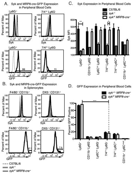
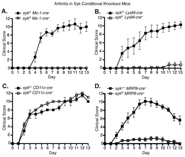
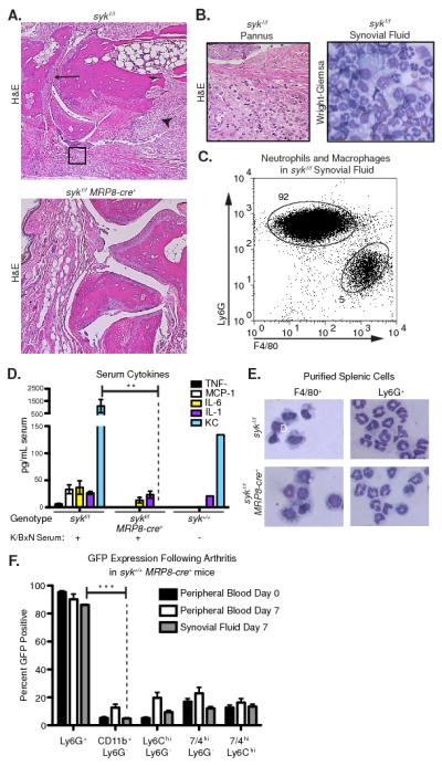
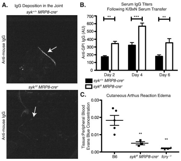
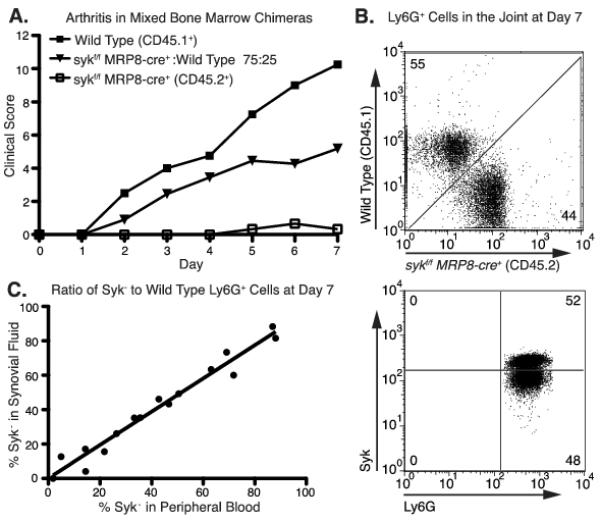
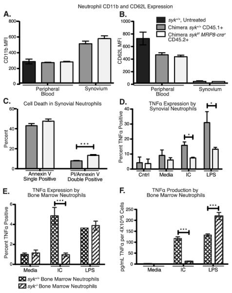
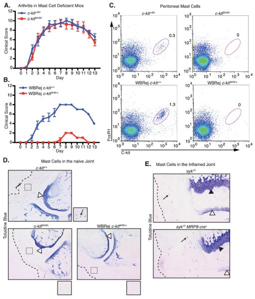
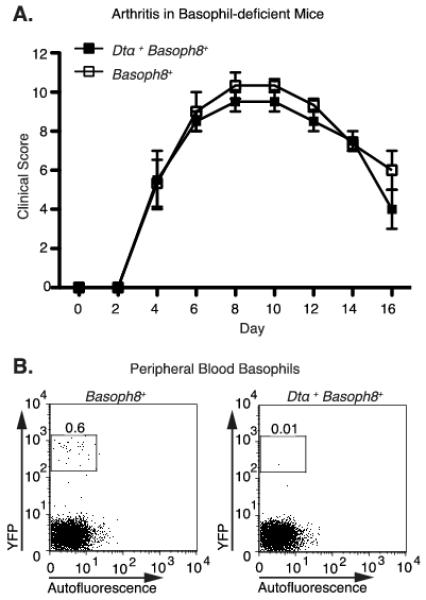
The K/BxN serum transfer model of arthritis is critically dependent on FcγR signaling events mediated by spleen tyrosine kinase (Syk). However, the specific cell types in which this signaling is required are not known. We report that deletion of Syk in neutrophils, achieved using syk(f/f) MRP8-cre(+) mice, blocks disease development in serum transfer arthritis. The syk(f/f) MRP8-cre(+) mice display absent joint disease and reduced deposition of pathogenic anti-glucose-6-phosphate isomerase Abs in the joint (with a reciprocal accumulation of these Abs in the peripheral circulation). Additionally, syk(f/f) MRP8-cre(+) mice manifest poor edema formation within 3 h after formation of cutaneous immune complexes (Arthus reaction). Together, this suggests that neutrophil-dependent recognition of immune complexes contributes significantly to changes in vascular permeability during the early phases of immune complex disease. Using mixed chimeric mice, containing both wild-type and syk(f/f) MRP8-cre(+) neutrophils, we find no impairment in recruitment of Syk-deficient neutrophils to the inflamed joint, but they fail to become primed, demonstrating lower cytokine production after removal from the joint. They also display an increased apoptotic rate compared with wild-type cells in the same joint. Mast cell-deficient c-kit(sh/sh) mice developed robust arthritis after serum transfer whereas c-kit(W/Wv) mice did not, suggesting that previous conclusions concerning the central role of mast cells in this model may need to be revised. Basophil-deficient mice also responded normally to K/BxN serum transfer. These results demonstrate that Syk-dependent signaling in neutrophils alone is critically required for arthritis development in the serum transfer model.


Figures

Figure 1
Figure 1
Neutrophil-specific deletion of Syk in sykf/f MRP8-cre+ mice. Peripheral blood and splenic leukocytes from mice of the indicated genotypes were stained for surface markers and Syk for analysis by flow cytometry, as described in the Materials and Methods. Cell populations were either analyzed for Syk or GFP expression. Leukocytes from syk−/− fetal liver chimeras served as a negative control for Syk staining. GFP positivity indicates the current expression of the MRP8-cre+-ires-GFP construct. A, B, Syk and GFP expression in peripheral blood and splenic leukocytes. Histograms are representative of at least 3 separate experiments, with at least 3 mice per genotype per experiment. C, Mean fluorescence intensity of Syk staining in peripheral blood populations in sykf/f MRP8-cre+ mice (n= 8) as compared to C57BL/6 wild type mice and sykf/f cells (n= 2-3). Only the significant differences by one-way ANOVA for Ly6G+ and 7/4hiLy6G cells are shown. Only staining in Ly6G+ cells was significantly different between sykf/f MRP8-cre+ and wild type controls. D, Percent GFP positive of peripheral blood leukocytes from syk+/+MRP8-cre+ and sykf/f MRP8-cre+ mice (n= 6). Only the significant differences, by one-way ANOVA, between Ly6G+ and 7/4hiLy6G cells in sykf/f MRP8-cre+mice are shown. GFP positivity in Ly6G+ cells was significantly higher than in all other cell populations analyzed. *=p<0.05, **=p<0.01, *** = p<0.001
Figure 2
Figure 2
Conditional deletion of Syk in myeloid cells or neutrophils specifically protects mice from K/BxN serum-induced arthritis. Clinical score was recorded daily following K/BxN serum transfer in mice of the indicated genotypes, as described in Materials/Methods. Solid squares represent littermate controls; hollow squares represent Syk conditional knockouts. A, sykf/f Mx-1-cre mice that have Syk deficiency in all hematopoietic cells (n = 3). B, sykf/−LysM-cre mice that have Syk deficiency in myeloid cells (n=4). C, sykf/f CD11c-cre mice that have Syk deficiency in dendritic cells (n=2). D, sykf/f MRP8-cre mice that have Syk deficiency in neutrophils (n=4). Representative plots from 2 - 6 independent experiments per genotype. Time points after day five were significant (p > 0.01, two-way ANOVA) between the conditional knockouts and wild type controls in panels A, B, and D.
Figure 3
Figure 3
Joint inflammation, synovial neutrophil recruitment and serum cytokine levels are elevated in sykf/f but not sykf/f MRP8-cre+ mice following K/BxN serum injection. A, Representative H&E-stained sections of sykf/f and sykf/f MRP8-cre+ forepaws 7 days after K/BxN serum transfer. Joints from sykf/f animals exhibit pannus formation and leukocyte infiltration (arrowhead), bone erosion (open arrowhead), and narrowing of the synovial space (arrow). B, Higher magnification view of sykf/f hindpaw (left) and Wright-Giemsa stain of isolated synovial fluid leukocytes (right). C, Synovial fluid leukocytes from sykf/f mice at Day 7 were stained with anti-Ly6G and F4/80 to enumerate neutrophils monocytes/macrophages, and analyzed by flow cytometry. D, The levels of the indicated cytokines in the serum of sykf/f and sykf/f MRP8-cre+ mice at 7 days following K/BxN serum transfer or untreated C57BL/6 mice was determined by luminex bead array as described in Materials and Methods. Data are from 4-10 mice per group and representative of two independent experiments. E, Splenocytes from sykf/f and sykf/f MRP8-cre+ mice were stained for F4/80 and Ly6G for sorting by flow cytometry to isolate monocytes/macrophages and neutrophils, respectively. Cytospun cells were then stained by Wright-Giemsa. F, Peripheral blood was collected from syk+/+MRP8-cre+ mice prior to K/BxN serum transfer (day 0, n=12) and day 7 following serum transfer (day 7, n=6). Synovial fluid was aspirated from the fore and hind-paws on day 7 (n=5). Peripheral and synovial leukocytes were stained as indicated and analyzed for GFP expression by flow cytometry. Ly6G+ cells were significantly more GFP positive than all other cell types (p > 0.01). Statistics analyzed by two-way ANOVA. **=p<0.01, *** = p<0.001
Figure 4
Figure 4
Neutrophil-specific deletion of Syk results in decreased joint antibody deposition following K/BxN serum transfer. A, Cryosections from the forepaws of syk+/+ MRP8-cre+ and sykf/f MRP8-cre+ mice 7 days following K/BxN serum transfer were stained for the Fc portion of IgG. Deposition of IgG lining the cartilage surface is shown by the arrow. B, Serum anti-GPI titers on days 2, 4, and 6 following K/BxN serum transfer in sykf/f and sykf/f MRP8-cre+ mice were determined by ELISA as described in Materials/Methods and analyzed by two-way ANOVA. C, Immune complex induced cutaneous edema was evaluated in syk+/+ MRP8-cre+, wild type, and FcγR−/− mice by Evans Blue extraction three hours following i.v. injection of antigen, as described in Materials and Methods. Results are shown as the ratio of Evans Blue concentration in the tissue to concentration in the peripheral blood (Statistics shown compared to C56BL/6 mice, by one-way ANOVA). **=p<0.01, *** = p<0.001
Figure 5
Figure 5
Syk-deficient neutrophils migrate equivalently to the inflamed joint. A, Mixed bone marrow chimeras were generated from sykf-f MRP8-cre+ (CD45.2+) and congenically marked wild type mice (C56BL/6; CD45.1+) in a ratios varying from 75% sykf/+MRP8-cre+ with 25% wild type, to 25% sykf/+MRP8-cre+. Control chimeras were also made with 100% wild type or sykf/f MRP8-cre+ bone marrow. Eight weeks following bone marrow transfer, chimeras were injected with K/BxN serum and clinical score was recorded as described in Material and Methods. Data shown are for control chimeras and the 75% sykf/fMRP8-cre+ mix only (n=5 mice per group). B, Synovial fluid neutrophils were isolated at day 7 following serum transfer from a mixed chimeric mouse (containing roughly 50% sykf/−MRP8-cre+ and 50% wild type cells) then stained for CD45.1, CD45.2 (top panel) and Syk protein (bottom panel). Peripheral blood neutrophils from a syk−/− chimeric mouse were used to define Syk negative cells (data not shown). C, Peripheral blood and synovial neutrophils from mixed chimeric mice were stained for Ly5.1 versus Ly5.2 to determine the percentage of sykf/−MRP8-cre+ versus wild type cells in each compartment as shown in B. The percentage of sykf/−MRP8-cre+ (designated % Syk-) in peripheral blood versus synovial fluid is shown for each individual mouse (R2 = 0.96).
Figure 6
Figure 6
Altered cytokine production, but not activation marker expression, by Syk-deficient neutrophils in the arthritic joint. A, B, Ly6G+ Neutrophils were isolated from the peripheral blood and synovial fluid of arthritic mixed bone marrow chimeras on day 7 following K/BxN serum transfer and stained for CD11b and CD62L (L-selectin). Mean fluorescence intensity of CD11b and CD62L for Syk-deficient (CD45.2+, white bars) or wild type (CD45.1+ gray bars) neutrophils are shown compared to peripheral blood neutrophils from a control B6 mouse not treated with serum (black bars). C, Day 7 synovial fluid neutrophils from mixed chimeric mice were stained with Ly6G, annexin V and propidium iodide (PI) and the percentage of positive cells for each marker are shown. A-C, n=7. D, Day 7 synovial fluid neutrophils were pooled from 8 mixed chimeric mice and stained for intracellular TNFα as described in Materials and Methods following 6 hours of incubation with media or media plus immune complexes (indicated as IC) or media plus 10 ng/ml LPS. Wild type versus sykf/f MRP8-cre+ cells were distinguished by CD45.1 versus CD45.2 staining. E, F, Naïve bone marrow neutrophils were isolated from either wild type or syk−/− fetal liver chimeric mice and stimulated in vitro with the indicated agonists for 6 hours, then E, stained for intracellular TNFα or F, culture supernatant was collected for TNFα ELISA as described in Materials and Methods (n = 3). Statistics analyzed by two-way ANOVA. IC = insoluble immune complex. *=p<0.05, **=p<0.01, *** = p<0.001
Figure 7
Figure 7
Mast cell deficiency does not affect the development or persistence of antibody-mediated arthritis. A, B, K/BxN serum was administered to c-kit+/Sh versus c-kitSh/Sh mice or wild-type versus c-kitW/W-v mice and disease was followed as described in Materials and Methods. C, Flow cytometric staining of resting peritoneal cells, stained for the mast cell markers C-kit and FcεR1 from c-kitSh/Sh, c-kitW/W-v and control c-kit+/+ or c-kit+/Sh mice. D, E, Toluidine blue staining as described in the Materials and Methods on D, forepaws from serum untreated c-kitSh/Sh, c-kitW/W-v and control mice, and E, hindpaws from sykf/f and sykf/f MRP8-cre+ mice 7 days after serum transfer. Magnified views of the regions highlighted by the squares are shown to the lower right of each panel. Dotted lines approximate the epidermis, arrows indicate mast cells, white arrowheads indicate cartilage, and black arrowheads indicate the epiphysial plate. Data shown are averaged from 3-5 mice per cohort.
Figure 8
Figure 8
Basophil deficiency does not affect the development or persistence of antibody-mediated arthritis. A, Clinical score was recorded following K/BxN serum injection in mice expressing a basophil-specific YFP-ires-cre (Basoph8+) either with or without the floxed Rosa-flxDTα (Dtα+) gene. B, Representative dot plots from Cre-expressing mice. Peripheral blood basophils were identified as CD4 autofluorescentlo YFP+ by flow cytometry.

References

    1. Borregaard N. Neutrophils, from marrow to microbes. Immunity. 2010;33:657–670. - PubMed
    1. Soehnlein O, Lindbom L. Phagocyte partnership during the onset and resolution of inflammation. Nat. Rev. Immunol. 2010;10:427–439. - PubMed
    1. Van Ziffle JA, Lowell CA. Neutrophil-specific deletion of Syk kinase results in reduced host defense to bacterial infection. Blood. 2009;114:4871–4882. - PMC - PubMed
    1. Cassatella MA, Locati M, Mantovani A. Never underestimate the power of a neutrophil. Immunity. 2009;31:698–700. - PubMed
    1. Cascao R, Rosario HS, Souto-Carneiro MM, Fonseca JE. Neutrophils in rheumatoid arthritis: More than simple final effectors. Autoimmun. Rev. 2010;9:531–535. - PubMed

Publication types

MeSH terms

Substances