

Inflammatory myofibroblastic tumor of the trachea with concomitant granulomatous lymph node lesions
- PMID: 21918650
- PMCID: PMC3171926
- DOI: 10.1155/2011/151729
Inflammatory myofibroblastic tumor of the trachea with concomitant granulomatous lymph node lesions
Abstract
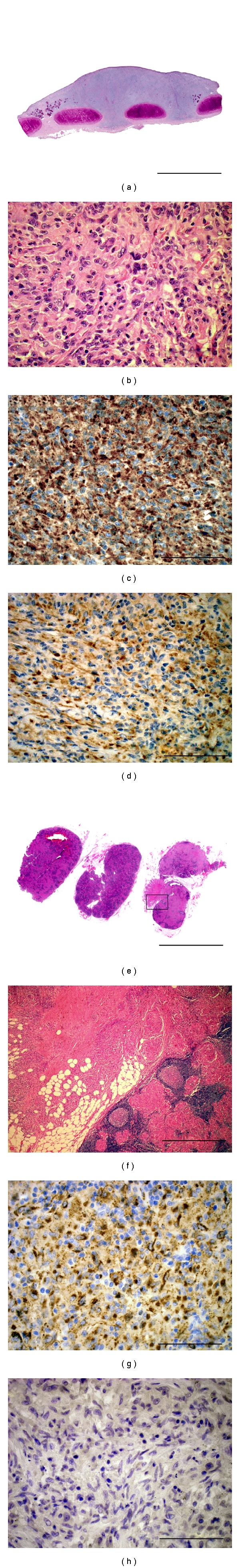
We report herein the case of a 57-year-old lady who had two concomittant lesions, an inflammatory myofibroblastic tumor in the trachea, and severe granulomatous lesions in the adjacent hilar lymph nodes. While these two lesions shared histological and some immunohistochemical features lesions. They differed in terms of ALK-1 expression, which was positive in the tracheal tumor and negative in the lymph nodes. The discussion of the case circles around putative pathophysiological links between the lesions. The authors favor the idea that the lymph nodes present a sarcoid-like granulomatous reaction to the inflammatory myofibroblastic tumor in the trachea over a coexistence of two independent entities. However, no conclusive evidence for this interpretation can be presented based on the existing literature.
Figures
Similar articles
-
Granulomatous slack skin mycosis fungoides developing simultaneously with sarcoid-like lesions in a patient with repeated anabolic injections in the past?Dermatol Ther. 2020 Jan;33(1):e13200. doi: 10.1111/dth.13200. Epub 2020 Jan 6. Dermatol Ther. 2020. PMID: 31854482
-
Inflammatory pseudotumor of lymph node and spleen: an entity biologically distinct from inflammatory myofibroblastic tumor.Hum Pathol. 2001 Dec;32(12):1382-7. doi: 10.1053/hupa.2001.29679. Hum Pathol. 2001. PMID: 11774173
-
Inflammatory pseudotumor of lymph nodes: a study of 25 cases with emphasis on morphological heterogeneity.Hum Pathol. 1997 Mar;28(3):332-8. doi: 10.1016/s0046-8177(97)90132-5. Hum Pathol. 1997. PMID: 9042798
-
Adult inflammatory myofibroblastic tumor of the trachea: case report and literature review.Respir Care. 2013 Jul;58(7):e72-6. doi: 10.4187/respcare.02198. Epub 2012 Dec 18. Respir Care. 2013. PMID: 23258581 Review.
-
Inflammatory myofibroblastic tumors of the lung carrying a chimeric A2M-ALK gene: report of 2 infantile cases and review of the differential diagnosis of infantile pulmonary lesions.Hum Pathol. 2017 Aug;66:177-182. doi: 10.1016/j.humpath.2017.06.013. Epub 2017 Jul 11. Hum Pathol. 2017. PMID: 28705706 Review.
Cited by
-
An Unusual Case of Systemic Inflammatory Myofibroblastic Tumor with Successful Treatment with ALK-Inhibitor.Case Rep Pathol. 2014;2014:470340. doi: 10.1155/2014/470340. Epub 2014 Jun 18. Case Rep Pathol. 2014. PMID: 25045570 Free PMC article.
-
Endotracheal inflammatory myofibroblastic tumour: A rare cause of central airway occlusion in adults.Respirol Case Rep. 2022 Jun 7;10(7):e0984. doi: 10.1002/rcr2.984. eCollection 2022 Jul. Respirol Case Rep. 2022. PMID: 35702693 Free PMC article.
References
-
- Ris HB, Krueger T, Cheng C, Pasche P, Monnier P, Magnusson L. Tracheo-carinal reconstructions using extrathoracic muscle flaps. European Journal of Cardio-Thoracic Surgery. 2008;33(2):276–283. - PubMed
-
- Coffin CM, Hornick JL, Fletcher CD. Inflammatory myofibroblastic tumor: comparison of clinicopathologic, histologic, and immunohistochemical features including ALK expression in atypical and aggressive cases. American Journal of Surgical Pathology. 2007;31(4):509–520. - PubMed
-
- Gleason BC, Hornick JL. Inflammatory myofibroblastic tumours: where are we now? Journal of Clinical Pathology. 2008;61(4):428–437. - PubMed
-
- Takeda SI, Onishi Y, Kawamura T, Maeda H. Clinical spectrum of pulmonary inflammatory myofibroblastic tumor. Interactive Cardiovascular and Thoracic Surgery. 2008;7(4):629–633. - PubMed
-
- Monfort-Gouraud M, Chokre R, Dubiez M, Ratsimihah T, Hamza A, Sauvageon G. Fait clinique: pseudotumeur inflammatoire de l’orbite et suspicion de sarcoidose. Archives de Pediatrie. 1996;3(7):697–700. - PubMed
Publication types
LinkOut - more resources
Full Text Sources
