. 2011;6(8):e24226.
doi: 10.1371/journal.pone.0024226. Epub 2011 Aug 30.

Helios expression is a marker of T cell activation and proliferation

Affiliations

Helios expression is a marker of T cell activation and proliferation

Tatiana Akimova et al. PLoS One. 2011.

Abstract

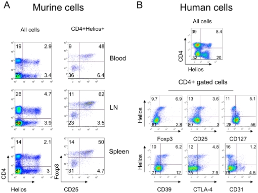
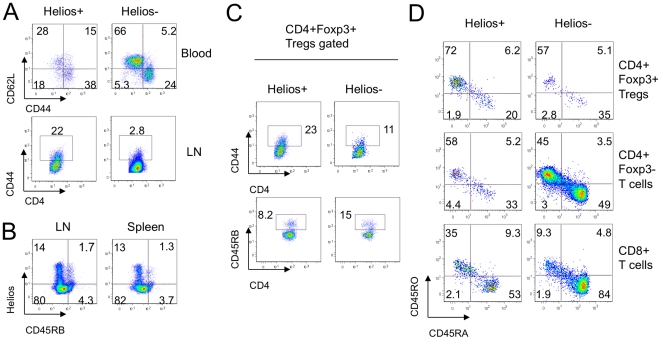
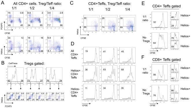
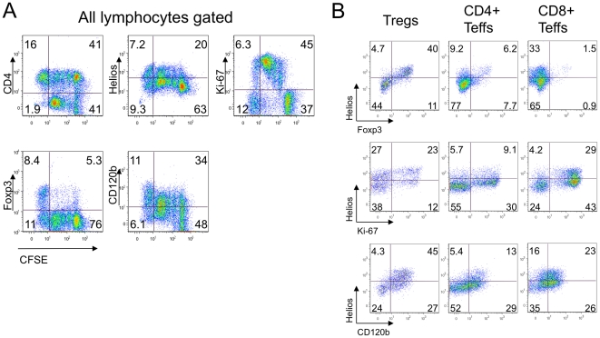
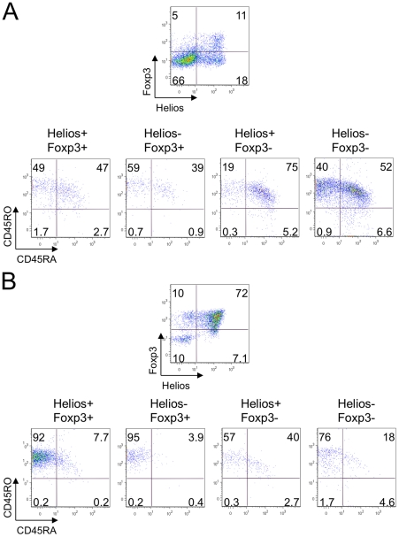
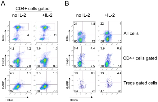
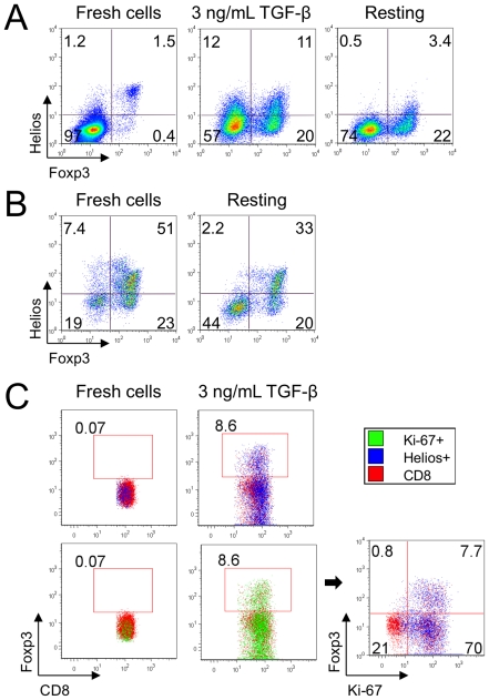
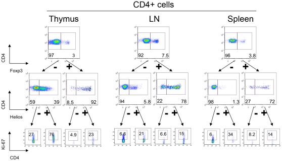
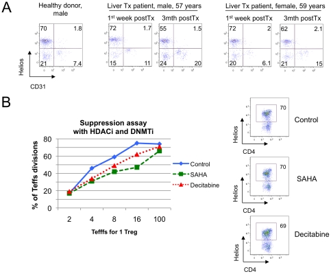
Foxp3+ T-regulatory cells (Tregs) normally serve to attenuate immune responses and are key to maintenance of immune homeostasis. Over the past decade, Treg cells have become a major focus of research for many groups, and various functional subsets have been characterized. Recently, the Ikaros family member, Helios, was reported as a marker to discriminate naturally occurring, thymic-derived Tregs from those peripherally induced from naïve CD4+ T cells. We investigated Helios expression in murine and human T cells under resting or activating conditions, using well-characterized molecules of naïve/effector/memory phenotypes, as well as a set of Treg-associated markers. We found that Helios-negative T cells are enriched for naïve T cell phenotypes and vice versa. Moreover, Helios can be induced during T cell activation and proliferation, but regresses in the same cells under resting conditions. We demonstrated comparable findings using human and murine CD4+Foxp3+ Tregs, as well as in CD4+ and CD8+ T cells. Since Helios expression is associated with T cell activation and cellular division, regardless of the cell subset involved, it does not appear suitable as a marker to distinguish natural and induced Treg cells.


Conflict of interest statement

Competing Interests: The authors have declared that no competing interests exist.

Figures

Figure 1
Figure 1. Helios expression was not restricted to CD4 cells, but among CD4+ cells correlated with Treg-associated markers.
(A) Expression of Helios in murine lymphocytes (left column) and CD25+ Foxp3+ co-expression in CD4+ Helios+ gated subsets (right column) of blood (top), lymph nodes (middle) and spleen (bottom). (B) Expression of Helios in human lymphocytes (top) and co-expression of Helios with Treg-associated markers in cells gated for CD4 expression. Right column, bottom row shows absence of correlation between Helios and CD31, a marker of recent thymic emigrants cells. Data are representative of 5 experiments.
Figure 2
Figure 2. Helios- cells appeared more naïve than Helios+ T cells.
(A) Expression of CD62L and CD44 in Helios+ (left column) and Helios- (right column) gated CD4+ cells in murine blood (top) and lymph nodes (second row). (B) Negative correlation between Helios and CD45RB expression in murine lymph nodes and spleen. (C) Expression of CD44 and CD45RB in Helios+ and Helios- gated Tregs in murine lymph nodes. (D) Maturation phenotype in human Tregs (top row), CD4+ Foxp3- Teffs (middle) and CD8+ T cells (bottom row) gated on Helios+ (left column) and Helios- (right column) subsets. Data are representative of 4 experiments.
Figure 3
Figure 3. Upregulation of Helios expression by Teff cells in suppression assays.
(A) Analysis of CD4+ gated murine Tregs and Teffs after 3 days of suppression assay showed that while Tregs gradually lose Foxp3 (top) and Helios (bottom) expression, a substantial proportion of Teff cells upregulate Helios (bottom), but not Foxp3 (top) expression; CFSE-negative gates show Tregs. (B) Treg division during suppression assays correlated with Helios and Foxp3 co-expression, while non-dividing Tregs lost both markers. Upper row: gating of dividing and non-dividing Tregs, with numbers showing % of Treg divisions at 1/1 (left), ½ (middle) and ¼ (right) ratios; arrows point to Helios and Foxp3 co-expression in corresponding divided and non-divided Tregs subsets through the same 1/1 to ¼ Treg/Teff ratios. (C) Tregs do not induce Helios expression in Teffs since CFSE+ CD4+ Teffs have similar Helios expression despite differing numbers of Tregs (1/1, ½ and ¼ ratios shown). (D) Analysis of Teffs gated into Helios+ and Helios- subsets showed Helios+ Teffs have a higher division rate and greater resistance to Treg suppression than Helios- mouse Teffs. Human Helios+ CD4+ (E) and Helios+ CD8 (F) Teffs showed higher divisions than Helios- cells regardless of Treg presence or absence, and Helios+ responder cells were more resistant to suppression than Helios- responders. Data are representative of 3 experiments.
Figure 4
Figure 4. Helios expression in suppression assay is associated with cell activation and division.
(A) After suppression assays, human Tregs and responders PBMC cells (1/2 Tregs/responders ratio) were stained for CD4, Helios, Ki-67, Foxp3 and CD120b; gating of live lymphocytes and co- expression of listed markers with CFSE divisions peaks shown, cells in CFSE-negative gate are Tregs. (B) Helios+ Tregs but not CD4+ and CD8+ responders showed co-expression of Helios and Foxp3 (top row). Helios+ cells showed more dividing (middle) and activated (bottom) phenotypes throughout all T cells subsets, including Tregs (left), CD4+ (middle) and CD8+ (right) cells. Data are representative of 2 experiments.
Figure 5
Figure 5. Helios is associated with an activated non-mature phenotype in suppression assays.
After suppression assays (1/2 Treg/responder ratio), human CD4+ Teffs (A) and Tregs (B) were divided into 4 subsets according to Helios and Foxp3 co-expression. Helios+Foxp3- cells had the least mature and the most activated phenotypes in human CD4+ Teffs (A) and Tregs (B) in suppression assays, while Foxp3 expressing cells were enriched for mature CD45RO+ CD45RA- subsets. Data are representative of 2 experiments.
Figure 6
Figure 6. IL-2 increased Helios expression in Ki-67+ Foxp3- GARP- cells.
Murine MNC from lymph nodes and spleen (A) and human PBMC (B) were stimulated with CD3/CD28±IL-2 for 24 hrs and stained for CD4, Foxp3, Helios, Ki-67 and GARP. IL-2 addition led to a slight increase in expression of Helios by Ki-67+ (A, top) CD4+ and CD4- (B, top) and Foxp3- cells (A, B, middle rows). Helios expression was not associated with GARP expression in CD4+ gated (A, bottom) or Foxp3+ gated (B, bottom) cells.
Figure 7
Figure 7. Helios is not associated with Treg lineage commitment in mice.
(A) Murine CD4+ CD25- Teffs stimulated with CD3/CD28 mAbs, IL-2 and TGF-beta to become iTregs upregulated Helios in both Foxp3+ and Foxp3- subsets (middle). Withdrawal of TCR stimulation and TGF-beta led to loss of Helios expression (right). (B) Murine Tregs after isolation (left) and after 4 d with IL-2 but without stimulation (right). In resting conditions Helios expression decreased, and Foxp3 expression by Tregs decreased mainly in the Helios+ subset. (C) Induction of Foxp3 in mouse CD8+ cells in the same conditions as in (A). Foxp3+ converted CD8 cells co-expressed Helios and Ki-67.
Figure 8
Figure 8. Helios+ expression correlates with Ki-67 expression in vivo.
Murine thymic (left), lymph node (middle) and splenic (right) CD4+ gated cells were divided into Foxp3+ Tregs and Foxp3- cells (top row), and then divided into Helios+ and Helios- subsets (middle), and Ki-67 expression within each subset was analyzed (bottom).
Figure 9
Figure 9. Helios expression in thymocytes depends on cell stimulation and not origin.
(A) Helios- Foxp3+ subsets were gated within murine thymic cells, and CD4 and CD8 distribution was analyzed. More than half (bottom) of Foxp3+ Helios- cells and 24% (top) of Foxp3hi Helios- cells in the thymus were immature CD4+ CD8+ DP thymocytes. (B) Murine thymic cells were incubated for 3 days with IL-2 only (non-stimulated, left column) or were stimulated with CD3/CD28 mAbs and IL-2 (stimulated, right column). Ki-67 expression (top) and Helios expression (middle) in non-stimulated cells decreased markedly; and Helios expression in thymic Tregs (CD4+ SP Foxp3+ cells) decreased from 91% to 57% (bottom).
Figure 10
Figure 10. Helios expression by human Tregs did not correlate with CD31 or suppressive function.
(A) Two liver transplant patients underwent serial analysis of Treg suppressive function, Foxp3 methylation and cell phenotype. At 3 mths post-transplant, Tregs had increased thymic output of CD31+ Tregs and reduction of Helios+ Tregs; Helios was not co-expressed with CD31. (B) Tregs, CD4+CD25- Teffs and CD4-depleted APC from healthy donor were used in suppression assays, with or without treatment with a DNMTi (Decitabine) or HDACi (SAHA). Enhancement of Treg suppression by either agent (left) was not accompanied by change in Helios expression (right).

References

    1. Vignali DA, Collison LW, Workman CJ. How regulatory T cells work. Nat Rev Immunol. 2008;8:523–532. - PMC - PubMed
    1. Feuerer M, Hill JA, Mathis D, Benoist C. Foxp3+ regulatory T cells: differentiation, specification, subphenotypes. Nat Immunol. 2009;10:689–695. - PubMed
    1. Hori S, Nomura T, Sakaguchi S. Control of regulatory T cell development by the transcription factor Foxp3. Science. 2003;299:1057–1061. - PubMed
    1. Fontenot JD, Gavin MA, Rudensky AY. Foxp3 programs the development and function of CD4+CD25+ regulatory T cells. Nat Immunol. 2003;4:330–336. - PubMed
    1. Pillai V, Ortega SB, Wang CK, Karandikar NJ. Transient regulatory T-cells: a state attained by all activated human T-cells. Clin Immunol. 2007;123:18–29. - PMC - PubMed

Publication types