

Molecular imaging with theranostic nanoparticles
- PMID: 21919457
- PMCID: PMC3196845
- DOI: 10.1021/ar200106e
Molecular imaging with theranostic nanoparticles
Abstract
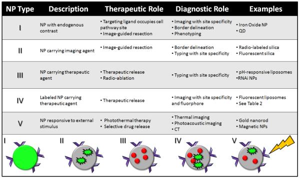
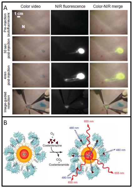
Nanoparticles (NPs) offer diagnostic and therapeutic capabilities not available with small molecules or microscale tools. As the field of molecular imaging has emerged from the blending of molecular biology with medical imaging, NP imaging is increasingly common for both therapeutic and diagnostic applications. The term theranostic describes technology with concurrent and complementary diagnostic and therapeutic capabilities. Although NPs have been FDA-approved for clinical use as transport vehicles for nearly 15 years, full translation of their theranostic potential is incomplete. However, NPs have shown remarkable success in the areas of drug delivery and magnetic resonance imaging. Emerging applications include image-guided resection, optical/photoacoustic imaging in vivo, contrast-enhanced ultrasound, and thermoablative therapy. Diagnosis with NPs in molecular imaging involves the correlation of the signal with a phenotype. The location and intensity of NP signals emanating from a living subject indicate the disease area's size, stage, and biochemical signature. Therapy with NPs uses the image for resection or delivery of a small molecule or RNA therapeutic. Ablation of the affected area is also possible via heat or radioactivity. The ideal theranostic NP includes several features: (1) it selectively and rapidly accumulates in diseased tissue; (2) it reports biochemical and morphological characteristics of the area; (3) it delivers an effective therapeutic; and (4) it is safe and biodegrades with nontoxic byproducts. Such a system contains a central imaging core surrounded by small molecule therapeutics. The system targets via ligands such as IgG and is protected from immune scavengers by a cloak of protective polymer. Although no NP has achieved all of the above criteria, many NPs possess one or more of these features. While the most clinically translatable NPs have been used in the field of magnetic resonance imaging, other types in development are quickly becoming more biocompatible through methods that modify their toxicity and biodistribution profiles. In this Account, we describe diagnostic imaging and therapeutic uses of NPs. We propose and offer examples of five primary types of nanoparticles with concurrent diagnostic and therapeutic uses.
Figures
References
-
- Massoud TF, Gambhir SS. Molecular imaging in living subjects: seeing fundamental biological processes in a new light. Genes Dev. 2003;17(5):545–580. - PubMed
-
- Weissleder R, et al. Molecular Imaging: Principles and Practice. People’s Medical Publishing House; Shelton, CT: 2010.
-
- Wagner V, et al. The emerging nanomedicine landscape. Nat. Biotechnol. 2006;24(10):1211–1218. - PubMed
-
- Kim BY, et al. Nanomedicine. N. Eng. J. Med. 2010;363(25):2434–43. - PubMed
-
- Sumer B, Gao J. Theranostic nanomedicine for cancer. Nanomedicine. 2008;3(2):137–140. - PubMed
Publication types
MeSH terms
Substances
Grants and funding
LinkOut - more resources
Full Text Sources
Other Literature Sources
Research Materials
Miscellaneous
