. 2012 Mar;18(5-6):520-32.
doi: 10.1089/ten.TEA.2011.0317. Epub 2011 Dec 2.

Dual regulation of breast tubulogenesis using extracellular matrix composition and stromal cells

Affiliations

Dual regulation of breast tubulogenesis using extracellular matrix composition and stromal cells

Silva Krause et al. Tissue Eng Part A. 2012 Mar.

Abstract

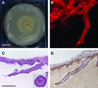
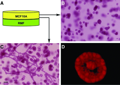
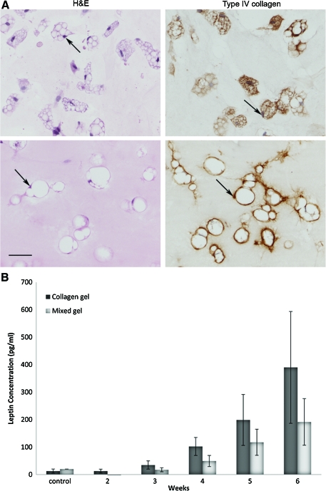
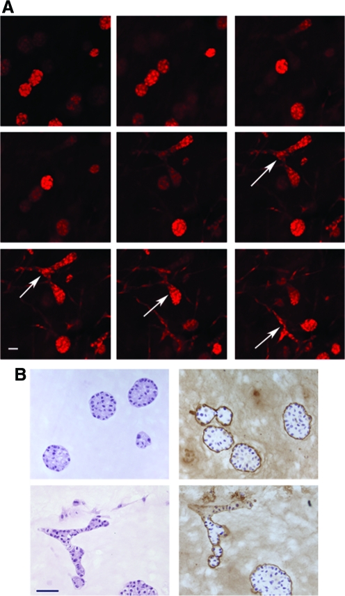
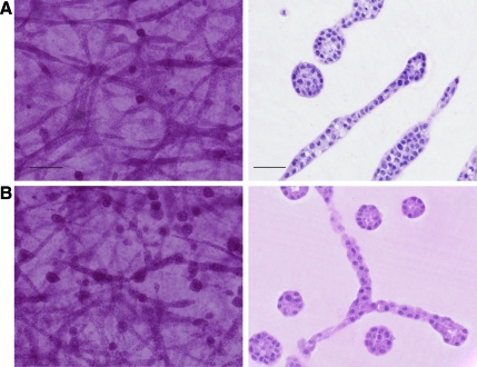
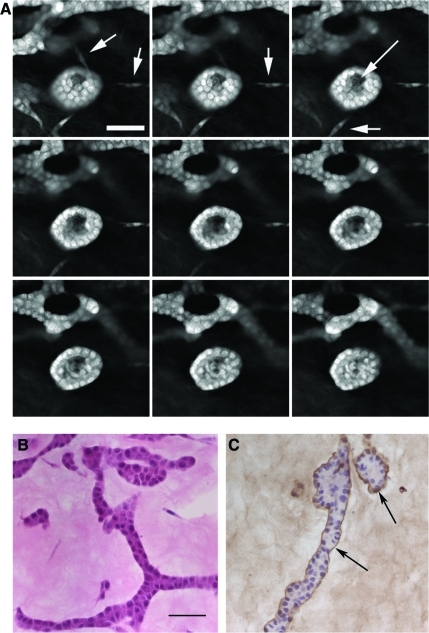
Epithelial-mesenchymal interactions during embryogenesis are critical in defining the phenotype of tissues and organs. The initial elongation of the mammary bud represents a central morphological event requiring extensive epithelial-mesenchymal crosstalk. The precise mechanism orchestrating this outgrowth is still unknown and mostly animal models have been relied upon to explore this process. Highly tunable three-dimensional (3D) culture models are a complementary approach to address the question of phenotypic determination. Here, we used a 3D in vitro culture to study the roles of stromal cells and extracellular matrix components during mammary tubulogenesis. Fibroblasts, adipocytes, and type I collagen actively participated in this process, whereas reconstituted basement membrane inhibited tubulogenesis by affecting collagen organization. We conclude that biochemical and biomechanical signals mediate the interaction between cells and matrix components and are necessary to induce tubulogenesis in vitro.


Figures

FIG. 1.
FIG. 1.
Experimental gel formats. Schematic depicts the following gel formats used in these studies: the basic three-dimensional gel (left), sandwich gel comprising two gels cast on top of each other with or without a porous membrane separating them (middle), and drop gels in which a drop of collagen–reconstituted basement membrane matrix is suspended within a collagen gel before solidification (right). Color images available online at www.liebertonline.com/tea
FIG. 2.
FIG. 2.
Epithelial structures obtained in mixed reconstituted basement membrane (rBM) –collagen gels containing MCF10A cells alone. Decreasing amounts of rBM (Matrigel) were used while the collagen concentration was maintained at 1 mg/mL in all cases. (A) Whole mounts (WM) and corresponding H&E were used to show the epithelial phenotype. Ductal structures (arrow) were first observed in the 20% rBM matrix. Ductal and alveolar structures were characterized using immunohistochemical analysis for basement membrane (laminin 5 staining) and polarization (sialomucin staining). Arrowheads indicate the presence of basement membrane surrounding ductal and alveolar structures. Diffuse staining is observed in the 5% rBM matrix. *Sialomucin expression at the apical surface of cells in an alveolar structure. Scale bar for WM, 200 μm; for histology, 100 μm. Time point: 2 weeks. (B) Morphometric analysis of epithelial structures in decreasing volumes of rBM in mixed gels (indicated in %) after 2 weeks in culture. *p<0.05, ** p<0.01. (C) Quantitative analysis of Ki67-positive epithelial cells in alveolar structures and in extending and tubular structures in decreasing volumes of rBM in mixed gels after 2 weeks in culture. *p<0.05. H&E, hematoxylin and eosin. Color images available online at www.liebertonline.com/tea
FIG. 3.
FIG. 3.
Picrosirius red staining and polarized light microscopy showing collagen fiber organization in gels containing MCF10A cells alone with decreasing volumes of reconstituted basement membrane (indicated in %). Scale bar: 50μm. Time point: 2 weeks. *Epithelial structures. Color images available online at www.liebertonline.com/tea
FIG. 4.
FIG. 4.
Presence of collagen was necessary for ductal formation. (A) Photograph of an acellular collagen gel containing a drop of MCF10A cell–containing mixed matrix (drop gel). (B): Confocal micrograph of the gel in (A) displaying a duct growing at the interface of the collagen gel and the reconstituted basement membrane (rBM) drop. (C) H&E staining of a duct growing at the interface of the collagen gel and an alveolar structure (inset) formed within the rBM drop. Alveolar and ductal structures have lumen, and the cells were polarized. (D) Laminin 5 staining indicating the presence of basement membrane surrounding the duct growing at the interface of the collagen gel. Time point: 2 weeks. Scale bars: (A) 5 mm, (C) 100 μm. Color images available online at www.liebertonline.com/tea
FIG. 5.
FIG. 5.
MCF10A cells and primary human fibroblasts obtained from reduction mammoplasties (RMF) need to be in close proximity for tubulogenesis to occur in a 50% reconstituted basement membrane (rBM)-based matrix. (A) Schematic representation of sandwich gel with RMF on bottom and MCF10A cells on top. The gels were separated by a porous membrane whose pore size allowed (3.0 μm) or prevented (0.22 μm) cell migration. (B) Whole-mounted MCF10A cell–containing gel in a plane distant from the RMF-containing gel. (C) Whole-mounted MCF10A cell–containing gel in a plane abutting the RMF-containing gel. (D) Confocal image of the top gel containing MCF10A cells showing lumen formation and polarized epithelial structures. Scale bar: 100 μm. Time point: 3 weeks. Color images available online at www.liebertonline.com/tea
FIG. 6.
FIG. 6.
(A) Human preadipocytes differentiated into adipocytes in collagen (top panel) and in a 50% reconstituted basement membrane (rBM) –based matrix (lower panel). Many of the adipocytes were unilocular and contained a displaced nucleus (arrows); some formed clusters containing a few cells, and secreted the basement membrane protein type IV collagen. Time point: 6 weeks. Scale bar: 50 μm. (B) Concentration of leptin (pg/mL) secreted by human adipocytes cultured in collagen or 50% rBM–based matrix sandwich gels. Conditioned medium from an acellular gel was used as a control for leptin production. Color images available online at www.liebertonline.com/tea
FIG. 7.
FIG. 7.
Proximity of adipocytes to MCF10A cells is necessary for tubulogenesis. (A) Consecutive confocal images of a z-stack arrangement of a MCF10A cell–containing gel cast on top of an adipocyte-containing mixed gel. Images on the upper left are farthest from the interface between the two different gel types, and those on the lower right are closest to the interface. MCF10A cells formed ductal structures only in the areas abutting the differentiated adipocyte-containing gel (arrow). Time point: 2 weeks. Scale bar: 20 μm. (B) Left panels: H&E of MCF10A cell–containing epithelial structures in the areas distant from (top panel) and abutting (bottom panel) the adipocyte-containing gel. Right panels: alveolar and ductal structures were characterized using immunohistochemical analysis for the presence of basement membrane (laminin 5 staining). Scale bar: 50 μm. Color images available online at www.liebertonline.com/tea
FIG. 8.
FIG. 8.
Human adipocytes did not affect the epithelial phenotype of (A) MCF10A cells alone in collagen gels or (B) MCF10A cells co-cultured with primary human fibroblasts obtained from reduction mammoplasties in 50% reconstituted basement membrane–based matrix. Carmine stained whole-mounted gels (left) and H&E staining of sections (right). Scale bars: whole-mounted: 200μm; H&E: 50μm. Color images available online at www.liebertonline.com/tea
FIG. 9.
FIG. 9.
Preadipocytes allow polarization and basement membrane and lumen formation. (A) Consecutive pictures of a confocal z-stack arrangement of a whole-mounted gel showing lumen formation (long arrow) when MCF10A cells were co-cultured with preadipocytes (short arrows) in a 50% reconstituted basement membrane (rBM)–based matrix. Time point: 2 weeks. Scale bar: 50 μm. (B) H&E of ductal structures obtained when preadipocytes were co-cultured with MCF10A in a 50% rBM–based matrix. Scale bar: 50 μm. (C) Laminin 5 staining indicates presence of a basement membrane (arrows) surrounding ductal and alveolar structures. Time point: 2 weeks. Color images available online at www.liebertonline.com/tea

Similar articles

Cited by

References

    1. Kratochwil K. Organ specificity in mesenchymal induction demonstrated in the embryonic development of the mammary gland of the mouse. Dev Biol. 1969;20:46. - PubMed
    1. Montesano R. Soriano J.V. Pepper M.S. Orci L. Induction of epithelial branching tubulogenesis in vitro. J Cell Physiol. 1997;173:152. - PubMed
    1. Sakakura T. Nishizuka Y. Dawe C.J. Mesenchyme-dependent morphogenesis and epithelium-specific cytodifferentiation in mouse mammary gland. Science. 1976;194:1439. - PubMed
    1. Cunha G.R. Role of mesenchymal-epithelial interactions in normal and abnormal development of the mammary gland and prostate. Cancer. 1994;74:1030. - PubMed
    1. Zangani D. Darcy K.M. Shoemaker S. Ip M.M. Adipocyte-epithelial interactions regulate the in vitro development of normal mammary epithelial cells. Exp Cell Res. 1999;247:399. - PubMed

Publication types

Substances

LinkOut - more resources