

Use of murine bioassay to resolve ovine transmissible spongiform encephalopathy cases showing a bovine spongiform encephalopathy molecular profile
- PMID: 21919992
- PMCID: PMC3505794
- DOI: 10.1111/j.1750-3639.2011.00526.x
Use of murine bioassay to resolve ovine transmissible spongiform encephalopathy cases showing a bovine spongiform encephalopathy molecular profile
Abstract
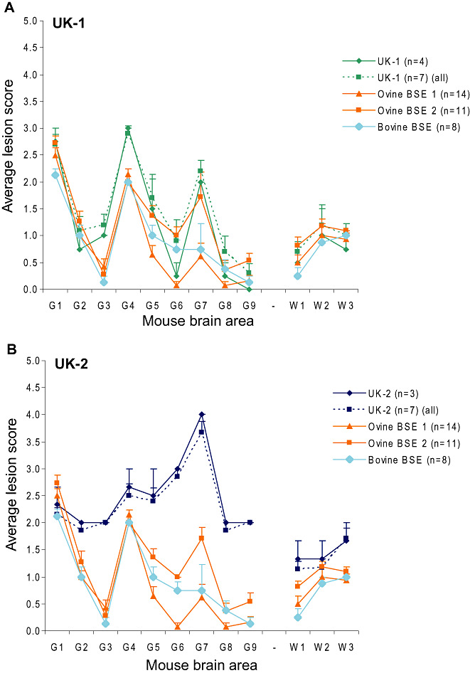
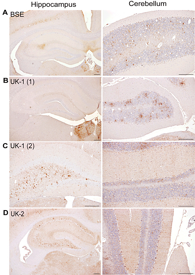
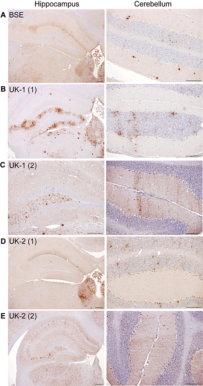
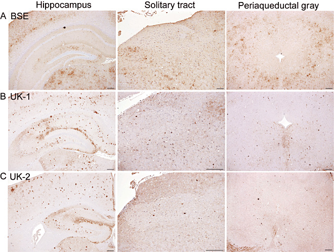
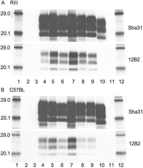
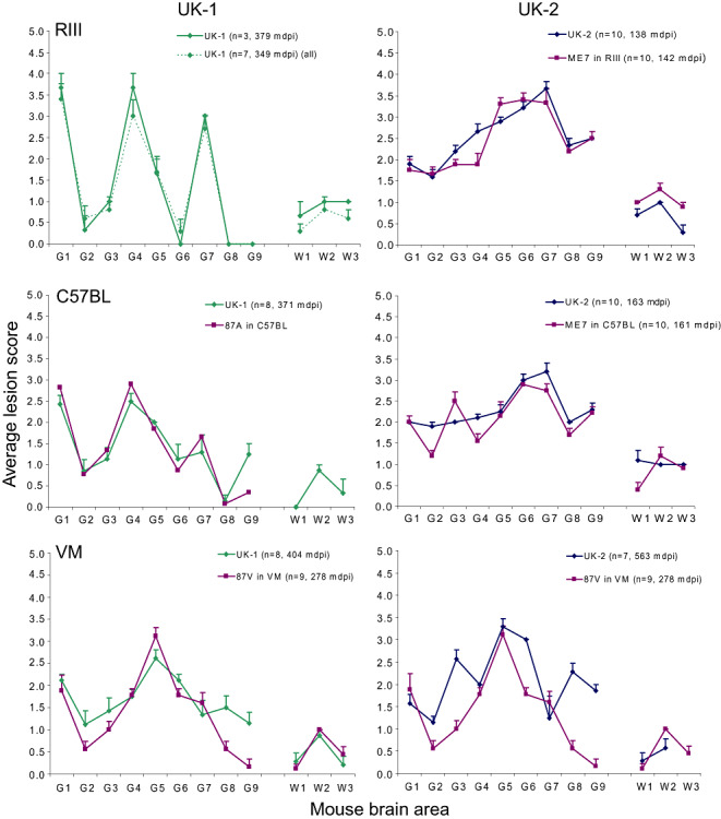
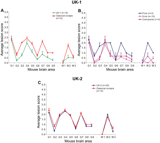
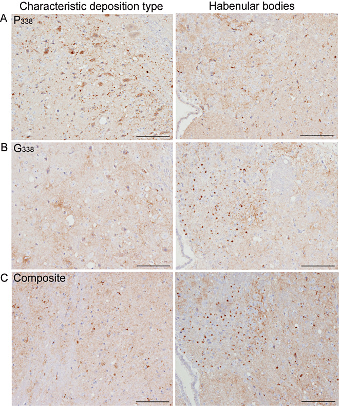
Two cases of unusual transmissible spongiform encephalopathy (TSE) were diagnosed on the same farm in ARQ/ARQ PrP sheep showing attributes of both bovine spongiform encephalopathy (BSE) and scrapie. These cases, UK-1 and UK-2, were investigated further by transmissions to wild-type and ovine transgenic mice. Lesion profiles (LP) on primary isolation and subpassage, incubation period (IP) of disease, PrP(Sc) immunohistochemical (IHC) deposition pattern and Western blot profiles were used to characterize the prions causing disease in these sheep. Results showed that both cases were compatible with scrapie. The presence of BSE was contraindicated by the following: LP on primary isolation in RIII and/or MR (modified RIII) mice; IP and LP after serial passage in wild-type mice; PrP(Sc) deposition pattern in wild-type mice; and IP and Western blot data in transgenic mice. Furthermore, immunohistochemistry (IHC) revealed that each case generated two distinct PrP(Sc) deposition patterns in both wild-type and transgenic mice, suggesting that two scrapie strains coexisted in the ovine hosts. Critically, these data confirmed the original differential IHC categorization that these UK-1 and UK-2 cases were not compatible with BSE.
© 2011 The Authors. Brain Pathology © 2011 International Society of Neuropathology.
Figures
Similar articles
-
Strain Typing of Classical Scrapie and Bovine Spongiform Encephalopathy (BSE) by Using Ovine PrP (ARQ/ARQ) Overexpressing Transgenic Mice.Int J Mol Sci. 2022 Jun 16;23(12):6744. doi: 10.3390/ijms23126744. Int J Mol Sci. 2022. PMID: 35743187 Free PMC article.
-
Further characterisation of transmissible spongiform encephalopathy phenotypes after inoculation of cattle with two temporally separated sources of sheep scrapie from Great Britain.BMC Res Notes. 2015 Jul 24;8:312. doi: 10.1186/s13104-015-1260-3. BMC Res Notes. 2015. PMID: 26205536 Free PMC article.
-
Transmission and characterization of bovine spongiform encephalopathy sources in two ovine transgenic mouse lines (TgOvPrP4 and TgOvPrP59).J Gen Virol. 2006 Dec;87(Pt 12):3763-3771. doi: 10.1099/vir.0.82062-0. J Gen Virol. 2006. PMID: 17098996
-
Pathology and pathogenesis of bovine spongiform encephalopathy and scrapie.Curr Top Microbiol Immunol. 2004;284:65-97. doi: 10.1007/978-3-662-08441-0_3. Curr Top Microbiol Immunol. 2004. PMID: 15148988 Review.
-
Insights into Mechanisms of Transmission and Pathogenesis from Transgenic Mouse Models of Prion Diseases.Methods Mol Biol. 2017;1658:219-252. doi: 10.1007/978-1-4939-7244-9_16. Methods Mol Biol. 2017. PMID: 28861793 Free PMC article. Review.
Cited by
-
Transmissibility of caprine scrapie in ovine transgenic mice.BMC Vet Res. 2012 Apr 2;8:42. doi: 10.1186/1746-6148-8-42. BMC Vet Res. 2012. PMID: 22472560 Free PMC article.
-
Selective propagation of mouse-passaged scrapie prions with long incubation period from a mixed prion population using GT1-7 cells.PLoS One. 2017 Jun 21;12(6):e0179317. doi: 10.1371/journal.pone.0179317. eCollection 2017. PLoS One. 2017. PMID: 28636656 Free PMC article.
-
Primary transmission of chronic wasting disease versus scrapie prions from small ruminants to transgenic mice expressing ovine or cervid prion protein.J Gen Virol. 2016 Sep;97(9):2451-2460. doi: 10.1099/jgv.0.000539. Epub 2016 Jul 8. J Gen Virol. 2016. PMID: 27393736 Free PMC article.
-
Re-transmissibility of mouse-adapted ME7 scrapie strain to ovine PrP transgenic mice.J Vet Sci. 2019 Mar;20(2):e8. doi: 10.4142/jvs.2019.20.e8. Epub 2019 Mar 12. J Vet Sci. 2019. PMID: 30944531 Free PMC article.
-
Incubation of ovine scrapie with environmental matrix results in biological and biochemical changes of PrP(Sc) over time.Vet Res. 2015 May 1;46(1):46. doi: 10.1186/s13567-015-0179-y. Vet Res. 2015. PMID: 25928902 Free PMC article.
References
-
- Aguzzi A (1998) Protein conformation dictates prion strain. Nat Med 4:1125–1126. - PubMed
-
- Beck KE, Sallis RE, Lockey R, Simmons MM, Spiropoulos J (2010) Ovine PrP genotype is linked with lesion profile and immunohistochemistry patterns after primary transmission of classical scrapie to wild‐type mice. J Neuropathol Exp Neurol 69:483–497. - PubMed
-
- Bellworthy SJ, Dexter G, Stack M, Chaplin M, Hawkins SAC, Simmons MM et al (2005) Natural transmission of BSE between sheep within an experimental flock. Vet Rec 157:206. - PubMed
Publication types
MeSH terms
Substances
LinkOut - more resources
Full Text Sources
Research Materials
