. 2011 Nov-Dec;32(10):1776-82.
doi: 10.3174/ajnr.A2634. Epub 2011 Sep 15.

Reducing patient radiation dose during CT-guided procedures: demonstration in spinal injections for pain

Affiliations

Reducing patient radiation dose during CT-guided procedures: demonstration in spinal injections for pain

T M Shepherd et al. AJNR Am J Neuroradiol. 2011 Nov-Dec.

Abstract

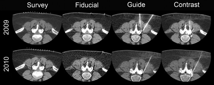
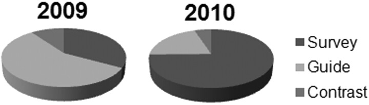
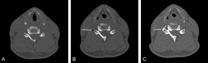
Background and purpose: CT guidance may improve precision for diagnostic and therapeutic spinal injections, but it can increase patient radiation dose. This study examined the impact of reducing tube current on patient radiation exposure and the technical success for these procedures, by using axial acquisitions for short scan lengths and eliminating nonessential imaging.

Materials and methods: Our institutional review board approved retrospective analysis of records from 100 consecutive outpatients undergoing spinal injections for pain before and after the CT protocol modification to reduce radiation dose. Data collected included patient age and sex, response to injection, number of sites and spinal levels treated, injection type, performing physician, CT acquisition method, number of imaging series, tube current, scan length, and DLP.

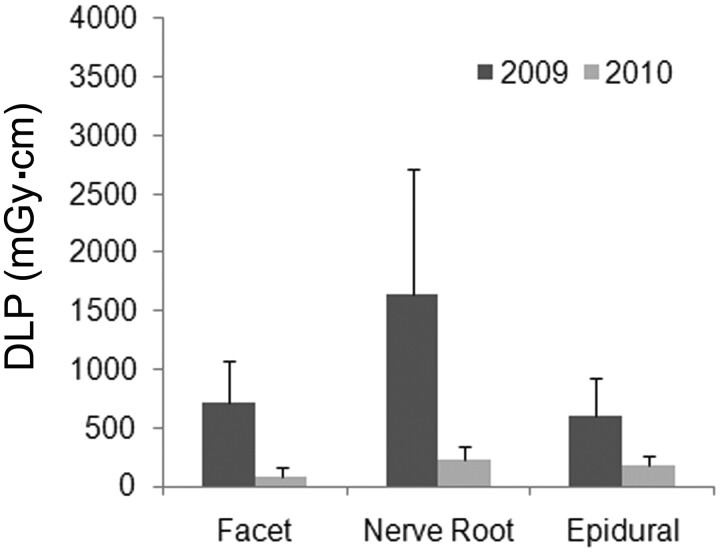
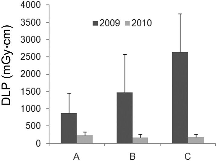
Results: Image contrast was reduced with the low-dose protocol, but this did not affect technical success or immediate pain relief. Mean DLP for all procedures decreased from 1458 ± 1022 to 199 ± 101 mGy · cm (P < .001). The range of radiologist-dependent DLP per procedure also was reduced significantly with the modified protocol. Selective nerve root blocks, lumbar injections, multiple injection sites, and the lack of prior imaging were each associated with a slightly higher DLP (<50 mGy · cm).

Conclusions: Radiation to patients undergoing CT-guided spinal injections can be decreased significantly without affecting outcome by reducing tube current, using axial acquisitions for short scan lengths, and eliminating nonessential imaging guidance. These measures also decrease variability in radiation doses between different practitioners and should be useful for other CT-guided procedures in radiology.


Figures

Fig 1.
Fig 1.
Comparison images for the same patient who received a left L4–5 facet block, a left L4–5 synovial cyst fenestration, and a left L4 selective nerve root block in both 2009 and 2010. All acquisitions in 2009 were helical, whereas in 2010, only the initial survey-phase CT images were acquired helically. The tube current was reduced from 549 to 149 mA in the survey image, from 84 to 30 mA in the fiducial bead image, from 84 to 50 mA in the guide phases of the study, and from 199 to 50 mA in the postcontrast images. Total examination DLP in 2010 was reduced 91%.
Fig 2.
Fig 2.
Between 2009 and 2010, there were significant reductions in DLP (milligrays per centimeter) for the total procedure as well as survey, guide, and contrast phases for all cases (A) and cervical-only (B) and lumbar-only (C) CT-guided spinal injections (P < .003 for all individual 2009 versus 2010 comparisons) (bar graph = mean ± standard error of the mean). The mean effective dose for cervical-only and lumbar-only CT-guided injections was reduced in 2010 from 9.7 to 1.1 mSv and from 17.5 to 3.3 mSv, respectively. DLP for some phases of the procedure also differed significantly within the same year (see “Results”).
Fig 3.
Fig 3.
Relative contributions from the survey, guide, and contrast phases of CT-guided spinal injections changed substantially between 2009 and 2010. Significant reductions in DLP during the guide phase of the examination in 2010 lowered its contribution to total procedure DLP from 57% to 20%. In 2010, almost 75% of the patient dose came from the initial survey CT images obtained for identifying anatomy relevant to the patient's spinal pain and prescribing the course of needle insertion.
Fig 4.
Fig 4.
Comparison of total examination doses for different CT-guided procedures that involved only 1 specific type of injection (facet joint, selective nerve root, or epidural block) (bar graph = mean ± SD). Between 2009 and 2010, the total dose decreased significantly for all 3 specific types of CT-guided spinal injections (ANOVA, P < .004 for all comparisons). Selective nerve root block procedures were consistently higher in total dose, but this trend only reached statistical significance compared with facet joint block procedures in 2010 (P = .004).
Fig 5.
Fig 5.
Dose reductions were observed for CT-guided spinal injections by individual supervising neuroradiologists (all 2009 versus 2010 comparisons, P < .003) (bar graph = mean ± SD). Comparisons between supervising neuroradiologists in the same year showed only trends toward statistically significant differences (ANOVA, P = .167 for either 2009 or 2010) due to the large variability in total DLP.
Fig 6.
Fig 6.
Lumbar-only procedures varied in the total number of sites (2.2 ± 1.3, 1–7) or levels (1.5 ± 1.0, 1–7) treated per encounter (mean ± SD, range), but the dose reduction observed persisted even when total examination dose (total DLP) was normalized for the number of sites (DLP/site) or levels (DLP/level) injected during the procedure (bar graph = mean ± SD). All 3 ways of analyzing procedural DLP showed approximately 80% reductions between 2009 and 2010 (P < .0001). Similar large dose reductions also persisted for cervical-only procedures when normalized for the number of sites or levels treated (P < .002).
Fig 7.
Fig 7.
Sample images from a 2010 study with aggressive dose reduction during a technically successful CT-guided right C6 selective nerve root block. Only the survey CT images were acquired helically (scan length = 50 mm). The milliampere/DLP (milligrays per centimeter) was 29/20.1, 10/0.5, and 20/1.0 for the survey (A), guide (B), and contrast (C) images, respectively (note, 8 guide series with slightly varying milliamperes or ranges were obtained). Total examination DLP was 28.6 mGy · cm, giving an estimated total effective dose of only 0.17 mSv. This dose is comparable with or lower than that for fluoroscopy-based spinal injections for pain.

References

    1. Gangi A, Dietemann JL, Mortazavi R, et al. . CT-guided interventional procedures for pain management in the lumbosacral spine. Radiographics 1998;18:621–33 - PubMed
    1. Deyo RA, Weinstein JN.. Low back pain. N Engl J Med 2001;344:363–70 - PubMed
    1. Brenner DJ, Hall EJ.. Computed tomography: an increasing source of radiation exposure. N Engl J Med 2007;357:2277–84 - PubMed
    1. Bogdanich W.. Radiation overdoses point up dangers of CT scans. New York Times. October 16, 2009;A13
    1. Smith-Bindman R, Lipson J, Marcus R, et al. . Radiation dose associated with common computed tomography examinations and the associated lifetime attributable risk of cancer. Arch Intern Med 2009;169:2078–86 - PMC - PubMed

Publication types