. 2011 Nov 11;286(45):39051-8.
doi: 10.1074/jbc.M111.274993. Epub 2011 Sep 15.

A three-part signal governs differential processing of Gli1 and Gli3 proteins by the proteasome

Affiliations

A three-part signal governs differential processing of Gli1 and Gli3 proteins by the proteasome

Erin K Schrader et al. J Biol Chem. .

Abstract

The Gli proteins are the transcriptional effectors of the mammalian Hedgehog signaling pathway. In an unusual mechanism, the proteasome partially degrades or processes Gli3 in the absence of Hedgehog pathway stimulation to create a Gli3 fragment that opposes the activity of the full-length protein. In contrast, Gli1 is not processed but degraded completely, despite considerable homology with Gli3. We found that these differences in processing can be described by defining a processing signal that is composed of three parts: the zinc finger domain, an adjacent linker sequence, and a degron. Gli3 processing is inhibited when any one component of the processing signal is disrupted. We show that the zinc fingers are required for processing only as a folded structure and that the location but not the identity of the processing degron is critical. Within the linker sequence, regions of low sequence complexity play a crucial role, but other sequence features are also important. Gli1 is not processed because two components of the processing signal, the linker sequence and the degron, are ineffective. These findings provide new insights into the molecular elements that regulate Gli protein processing by the proteasome.


Figures

FIGURE 1.
FIGURE 1.
Gli3 processing is appropriately regulated in HEK293T cells, and the Gli3 PDD does not induce Gli1 processing. In this and all subsequent figures, HEK293T cells were transfected with N-terminally FLAG-tagged Gli1 or Gli3 and constitutively active PKA, unless otherwise indicated. Gli1 and Gli3 expression and processing were assessed by immunoblotting with a FLAG antibody after cell lysis and SDS-PAGE. Results in this and all subsequent figures are representative of at least three independent experiments. A, schematic diagrams of Gli1 (top) and Gli3 (bottom). The zinc fingers are depicted as small rectangles, the PKA phosphorylation sites are shown as asterisks, and the FLAG tag is depicted as a green rectangle. The FLAG tag is omitted from all other figures for clarity. B, Gli3 was processed and Gli1 was not processed in HEK293T cells. The processed fragment sometimes appears as a doublet in other investigations of Ci/Gli3 processing as well (5, 6, 17, 24). Gli3 processing was dependent on the proteasome; addition of the proteasome inhibitor lactacystin (Lac) abolished processing. C, Gli3 processing was dependent on PKA activity. Activation of PKA, either through the addition of forskolin (FSK) or co-transfection of constitutively active PKA, induced Gli3 processing. Mutation of the first four PKA sites in Gli3 (Gli3(Mut PKA)) abolished processing. Mock, cells transfected with empty pcDNA3. D, in this and all subsequent figures, schematic diagrams of the constructs are shown in the order top to bottom that they are depicted left to right in the blot, and the first construct in each set of diagrams is wild-type Gli1 or Gli3. Gli1 containing the Gli3 processing determinant domain (24) (2. Gli1(PDD Gli3)) was not processed. Gli1 containing the Gli3 zinc fingers and C terminus was processed (3. Gli1(C term Gli3)). E, the processing signal in Gli3. The features between the zinc finger domain (ZFs, small rectangles) and the PKA site region (asterisks) are enlarged. The two simple sequence regions are labeled as 1 and 2. The four lysines of the Gli3 processing degron, the previously defined processing determinant domain (PDD) (24), and the predicted end of the processed form of Gli3 (19) are indicated. The components of the processing signal, as we define them, are indicated below the diagram.
FIGURE 2.
FIGURE 2.
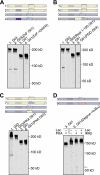
Gli3 processing requires a folded domain. A, replacement of all five zinc fingers in Gli3 with an unstructured region from S. cerevisiae cytochrome b2 (red) abolished processing (2. Gli3(ZnFUnstruc)). B, replacement of the zinc fingers in Gli3 with E. coli DHFR (purple) enhanced processing (3. Gli3(ZnFDHFR)). Lac, lactacystin.
FIGURE 3.
FIGURE 3.
A linker sequence adjacent to the zinc fingers is required for Gli3 processing. The linker sequence, which is the 140 amino acids between the zinc fingers and the processing degron, is enlarged in the diagrams. A, Gli3 processing was significantly impaired when the first 100 amino acids after the zinc fingers were replaced with a complex, unstructured region from S. cerevisiae cytochrome b2 (red, 2. Gli3(Repl 6–100)). Deletion of the endogenous simple sequences in Gli3 (blue, 3. Gli3(ΔSimple)) or replacement of both simple sequences with a complex sequence (red, 4. Gli3(SimpleComplex)) also eliminated Gli3 processing. When the simple sequences in Gli3 were replaced with an exogenous simple sequence, the glycine-rich region from p105 (green), processing was induced that was dependent on PKA activity and the proteasome (5. Gli3(SimpleGRR)). B, replacement of the entire 140-amino acid linker sequence with a complex, unstructured sequence (red) containing an insertion of a very low complexity sequence (35 glycines, green) abolished Gli3 processing (6. Gli3(Repl 6–140+polyG)). Replacement of amino acids 101–140 after the zinc fingers with a complex, unstructured sequence (red) also disrupted processing (7. Gli3(Repl 101–140)). Lac, lactacystin.
FIGURE 4.
FIGURE 4.
The location, but not the identity, of the Gli3 degron is critical for processing. A, transfer of the entire Gli3 degron (the lysines (tan) and the PKA site region (brown)) to the C terminus of Gli3 abolished processing (2. Gli3(DegronC term)), even when a copy of the lysines was left at the endogenous location (3. Gli3(Lys+DegronC term)). B, replacement of the Gli3 PKA site region with the IκBα degron (pink) permitted processing (4. Gli3(DegronIκBα)). The IκBα degron is activated by IKK (40, 41, 51, 52). Endogenous IKK appears to be active at a low level in HEK293T cells, and further IKK activity can be induced by overexpression of activated IKKβ. Lac, lactacystin.
FIGURE 5.
FIGURE 5.
Enhancing each component of the Gli processing signal individually is not sufficient to induce robust Gli1 processing. A, the Gli1 zinc fingers were sufficient to mediate processing in the context of Gli3 (2. Gli3(ZnF Gli1)). Replacement of the Gli1 zinc fingers with E. coli DHFR (purple, 4. Gli1(ZnFDHFR)) did not cause Gli1 processing. B, Gli3 processing was significantly impaired when the first 100 amino acids after the zinc fingers were replaced with the analogous sequence from Gli1 (light blue, 5. Gli3(Repl 1–100 Gli1)). The previously defined Gli3 PDD (24) (6. Gli1(PDD Gli3)) did not induce Gli1 processing. The Gli3 PDD contains the Gli3 linker sequence and the lysines of the processing degron. C, swapping the Gli1 PKA site region (light blue) into Gli3 inhibited Gli3 processing (7. Gli3(PKA Gli1)). Swapping the Gli3 PKA site region (yellow) into Gli1 did not cause Gli1 processing (8. Gli1(PKA Gli3)). D, replacement of the Gli1 PKA site region with the IκBα degron (pink) induced very weak Gli1 processing when the IκBα degron was stimulated with constitutively active IKKβ (9. Gli1(DegronIκBα)). The processed fragment is indicated with an arrow. Lac, lactacystin.
FIGURE 6.
FIGURE 6.
Changing two processing signal components in combination induces Gli1 processing. A, the Gli3 PDD (yellow) caused robust Gli1 processing when the Gli1 PKA site region was also replaced with the IκBα degron (pink, 3. Gli1(DegronIκBα+Gli3 PDD)). Gli1 with only the IκBα degron is shown for comparison (2. Gli1(DegronIκBα)). B, Gli1 containing both the Gli3 linker sequence and most of the Gli3 degron (yellow) was processed (4. Gli1(Repl 1–314 Gli3)). C, when the Gli1 zinc fingers were replaced with a domain more resistant to unfolding (DHFR, purple) and a simple sequence (glycine-rich region, green) was inserted C-terminal to zinc fingers, Gli1 was processed (7. Gli1(ZnFDHFR+GRR)). Neither the insertion of DHFR (5. Gli1(ZnFDHFR)) nor the simple sequence alone (6. Gli1(GRR)) led to Gli1 processing. IKKβ, constitutively active IKKβ; Lac, lactacystin.

Similar articles

Cited by

References

    1. Hooper J. E., Scott M. P. (2005) Nat. Rev. Mol. Cell Biol. 6, 306–317 - PubMed
    1. Jiang J., Hui C. C. (2008) Dev. Cell 15, 801–812 - PMC - PubMed
    1. Kent D., Bush E. W., Hooper J. E. (2006) Development 133, 2001–2010 - PubMed
    1. Zhang Q., Zhang L., Wang B., Ou C. Y., Chien C. T., Jiang J. (2006) Dev. Cell 10, 719–729 - PubMed
    1. Aza-Blanc P., Ramírez-Weber F. A., Laget M. P., Schwartz C., Kornberg T. B. (1997) Cell 89, 1043–1053 - PubMed

Publication types

MeSH terms

LinkOut - more resources