. 2011 Oct;22(10):1809-14.
doi: 10.1681/ASN.2011010084. Epub 2011 Sep 15.

Macrophages promote cyst growth in polycystic kidney disease

Affiliations

Macrophages promote cyst growth in polycystic kidney disease

Anil Karihaloo et al. J Am Soc Nephrol. 2011 Oct.

Abstract

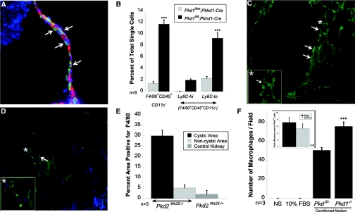
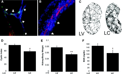
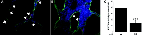
Polycystic kidney disease (PKD) exhibits an inflammatory component, but the contribution of inflammation to cyst progression is unknown. Macrophages promote the proliferation of tubular cells following ischemic injury, suggesting that they may have a role in cystogenesis. Furthermore, cultured Pkd1-deficient cells express the macrophage chemoattractants Mcp1 and Cxcl16 and stimulate macrophage migration. Here, in orthologous models of both PKD1 and PKD2, abnormally large numbers of alternatively activated macrophages surrounded the cysts. To determine whether pericystic macrophages contribute to the proliferation of cyst-lining cells, we depleted phagocytic cells from Pkd1(fl/fl);Pkhd1-Cre mice by treating with liposomal clodronate from postnatal day 10 until day 24. Compared with vehicle-treated controls, macrophage-depleted mice had a significantly lower cystic index, reduced proliferation of cyst-lining cells, better-preserved renal parenchyma, and improved renal function. In conclusion, these data suggest that macrophages home to cystic areas and contribute to cyst growth. Interruption of these homing and proliferative signals could have therapeutic potential for PKD.


Figures

Figure 1.
Figure 1.
Macrophages surround cysts in murine models of PKD. Kidneys were perfusion fixed with 4% PFA and cryosections stained for macrophage marker F4/80. (A) Cyst-lining cells showing closely apposed macrophages stained green. Green, F4/80; Red, DBA. (B) Quantitative analysis of FACS-sorted macrophages defined as F4/80+CD45+CD11c- cells and further analyzed for Ly6c expression; ***P < 0.0001. (C) Cystic area from Pkd2WS25/- kidney showing several F4/80+ cells (green). Inset shows magnified view of area marked with an asterisk. (D) Noncystic area from Pkd2ws25/+ kidney showing very few F4/80+ cells. Inset shows magnified view of selected area indicated by the asterisk. (E) Quantitative analysis of macrophages in kidneys from Pkd2WS25/- and Pkd2ws25/+ (control) mice. (F) Transwell migration of macrophages toward DMEM-F12, DMEM-F12 + 10%FBS, DMEM-F12 conditioned medium from Pkd1fl/- cells, and DMEM-F12 conditioned media from Pkd1-/- cells, respectively, n = 3. Inset: qRT-PCR for Mcp-1 and Cxcl16 was performed on RNA from Pkd1fl/- and Pkd1-/- cells and reported as the fold increase of Pkd1-/- as compared with Pkd1fl/-; n = 3.
Figure 2.
Figure 2.
Clodronate treatment reduces macrophage load and improves renal function. Cystic kidneys from Pkd1fl/fl;Pkhd1-Cre mice treated with LC show (B) fewer F4/80+ macrophages (arrows) along the cyst-lining cells compared with LV-treated mouse kidneys (A, arrows). (C) Scan of a sagittal section of a representative kidney from LV- and LC-treated mice, respectively, shows (D) better-preserved parenchyma in LC-treated mice and lowered cystic index, (E) improved kidney/body weight ratio, and (F) significantly improved renal BUN values. A, B: Green, F4/80; Red, DBA. *P < 0.01; **P < 0.001.
Figure 3.
Figure 3.
LC administration reduces cystic-lining cell proliferation. Representative kidney sections are shown from (A) LV-treated and (B) LC-treated mice stained for Ki67 (arrows). Green, DBA, Red/purple-Ki67; Blue, DAPI. (C) Ki67 positive nuclei quantitated within DBA positive tubules from kidneys of LV- and LC-treated mice. Data pooled from eight individual mice is shown. ***P < 0.0001.

References

    1. Gallagher AR, Germino GG, Somlo S: Molecular advances in autosomal dominant polycystic kidney disease. Adv Chronic Kidney Dis 17, 118–130, 2010 - PMC - PubMed
    1. Torres VE, Harris PC: Autosomal dominant polycystic kidney disease: The last 3 years. Kidney Int 76, 149–168, 2009 - PMC - PubMed
    1. Harris PC, Torres VE: Polycystic kidney disease. Annu Rev Med 60, 321–337, 2009 - PMC - PubMed
    1. Nadella R, Blumer JB, Jia G, Kwon M, Akbulut T, Qian F, Sedlic F, Wakatsuki T, Sweeney WE, Jr, Wilson PD, Lanier SM, Park F: Activator of G protein signaling 3 promotes epithelial cell proliferation in PKD. J Am Soc Nephrol 21, 1275–1280, 2010 - PMC - PubMed
    1. Gardner KD, Jr, Burnside JS, Elzinga LW, Locksley RM: Cytokines in fluids from polycystic kidneys. Kidney Int 39, 718–724, 1991 - PubMed

Publication types

LinkOut - more resources