. 2011 Nov;22(11):2004-15.
doi: 10.1681/ASN.2010121249. Epub 2011 Sep 15.

Hypoxia-inducible transcription factors stabilization in the thick ascending limb protects against ischemic acute kidney injury

Affiliations

Hypoxia-inducible transcription factors stabilization in the thick ascending limb protects against ischemic acute kidney injury

Gunnar Schley et al. J Am Soc Nephrol. 2011 Nov.

Abstract

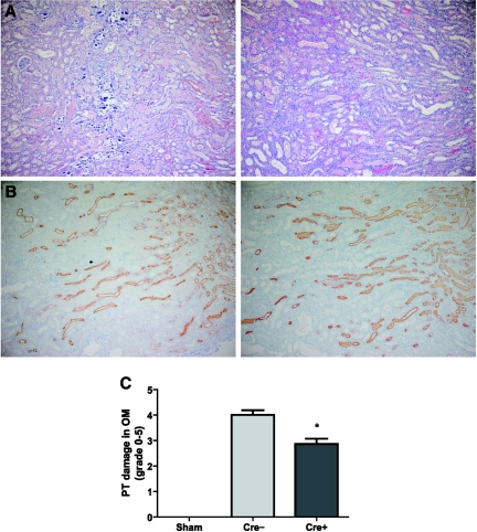
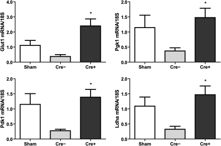
Hypoxia-inducible transcription factors (HIF) protect cells against oxygen deprivation, and HIF stabilization before ischemia mitigates tissue injury. Because ischemic acute kidney injury (AKI) often involves the thick ascending limb (TAL), modulation of HIF in this segment may be protective. Here, we generated mice with targeted TAL deletion of the von Hippel-Lindau protein (Vhl), which mediates HIF degradation under normoxia, using Tamm-Horsfall protein (Thp)-driven Cre expression. These mice showed strong expression of HIF-1α in TALs but no changes in kidney morphology or function under control conditions. Deficiency of Vhl in the TAL markedly attenuated proximal tubular injury and preserved TAL function following ischemia-reperfusion, which may be partially a result of enhanced expression of glycolytic enzymes and lactate metabolism. These results highlight the importance of the thick ascending limb in the pathogenesis of AKI and suggest that pharmacologically targeting the HIF system may have potential to prevent and mitigate AKI.


Figures

Figure 1.
Figure 1.
Generation of mice deficient for Vhl in thick ascending limbs of Henle's loop. (A) Scheme of mouse Tamm-Horsfall protein (Thp) promoter Cre-recombinase DNA construct. (B) Detection of Cre mRNA by RT-PCR in different organs of mice heterozygous for the Thp-Cre construct. The gel is representative of results obtained from offsprings derived from line 27 Thp-Cre mice. (C) RT-PCR analysis of genomic DNA isolated from different organs of Thp-Cre+;Vhl+f/+f mice with primers amplifying the nonrecombined (2-lox) and the recombined (1-lox) Vhl conditional allele. Lanes were run on the same gel but were noncontiguous. (D) Deletion efficiency of the loxP-flanked Vhl allele (Cre+; n = 3) in microdissected TAL cells was determined by real-time PCR in comparison to the WT allele (Cre−; n = 3). Representative consecutive kidney sections (magnification from the corticomedullary junction) displaying immunohistochemistries for HIF-1α (E) and THP (F) of 3-month-old Vhl knockout mice. Original magnification, ×400.
Figure 2.
Figure 2.
Diuretic response to furosemide was not affected by Vhl deficiency. Urine osmolality (A) and urine flow rates (B) after intraperitoneal administration of 40 mg/kg furosemide in WT (Cre−; n = 19) and Vhl knockout mice (Cre+; n = 16).
Figure 3.
Figure 3.
Loss of Vhl in TAL cells induced HIF target genes and increased anaerobic glucose utilization. (A) mRNA expression of HIF target genes Glut1, Pgk1, Pdk1, and Ldha in microdissected proximal convoluted tubules (PCT) and TALs of WT (Cre−; n = 4) and Vhl deficient mice (Cre+; n = 7) analyzed by real-time PCR. Lactate (B) and ATP (C) concentrations in total kidney tissue of WT (Cre−; n = 9) and Vhl knockout animals (Cre+; n = 9) after abdominal laparotomy (left panel) and 5 minutes after warm ischemia (right panel), simulated by incubation in 37°C water bath. *, P < 0.05, Cre− versus Cre+.
Figure 4.
Figure 4.
Kidney histomorphology was significantly ameliorated in knockout mice after renal ischemia-reperfusion. Representative microphotographs of H&E staining (A) and immunostaining for THP (B) in kidney sections (magnification from outer medulla) of WT (left) and Vhl deficient mice (right) 3 days after ischemia-reperfusion. Note that the calcification was present only in the WT group. Original magnification, ×100. (C) Semiquantitative analysis of proximal tubular (PT) damage in outer medulla (OM) of WT (Cre−; n = 18) and Vhl knockout mice (Cre+; n = 22) in comparison to sham-operated Cre− (n = 4) and Cre+ mice (n = 4). *, P < 0.05, Cre− versus Cre+.
Figure 5.
Figure 5.
Kidney function was significantly improved in knockout mice after renal ischemia-reperfusion. (A) Plasma creatinine (left panel) and urea (right panel) 3 days after kidney ischemia in WT (Cre−; n = 19) and Vhl knockout mice (Cre+; n = 19). Both sham-operated WT (n = 3) and Vhl deficient mice (n = 3) served as controls. (B) mRNA expression of the renal biomarkers Ngal (left) und Kim1 (right) measured by real-time PCR in postischemic total kidney extracts of WT (n = 5) and Vhl deficient mice (n = 5) in comparison to sham-operated Cre− (n = 3) and Cre+ mice (n = 2). (C) Time course of urine osmolality after intraperitoneal administration of 40 mg/kg furosemide in WT (Cre−; n = 5) and Vhl deficient (Cre+; n = 5) mice 3 days after renal ischemia-reperfusion. (D) Proliferating cells per TAL cross section in postischemic WT (Cre−; n = 8) and Vhl knockout kidneys (Cre+; n = 8) as well as sham-operated Cre− (n = 2) and Cre+ kidneys (n = 2). Immunhistochemistry for PCNA and THP was performed on consecutive sections. (E) Tubular apoptosis in the outer medulla of postischemic WT (Cre−; n = 5) and knockout mice (Cre+; n = 5) assessed by TUNEL staining. Sham-operated Cre− (n = 2) and Cre+ kidneys (n = 1) served as controls. *, P < 0.05, Cre− versus Cre+.
Figure 6.
Figure 6.
The inflammatory reaction in postischemic kidneys was not affected by Vhl deletion. (A) Semiquantitative analysis of inflammatory cell infiltration in H&E stained kidney sections of WT (Cre−) and Vhl knockout (Cre+) mice after renal ischemic (Cre− n = 18, and Cre+ n = 22) or sham (Cre− n = 4, and Cre+ n = 4) surgeries. (B) Infiltration with F4/80-positive macrophages in postischemic kidneys of WT (Cre−; n = 6) and Vhl deficient mice (Cre+; n = 6) as well as sham-operated Cre− (n = 2) and Cre+ mice (n = 2). (C) mRNA levels of the inflammatory cytokines Tnfα, Il1ß, Il6, and Mcp1 in total kidney extracts of WT (Cre−; n = 5) and Vhl knockout mice (Cre+; n = 5) as measured by real-time PCR. Sham-operated Cre− (n = 2) and Cre+ kidneys (n = 2) served as controls.
Figure 7.
Figure 7.
Expression of glycolytic enzymes was preserved in postischemic kidneys of knockout mice. Relative mRNA expression of the glycolytic enzymes Glut1, Pgk1, Pdk1, and Ldha in total kidney extracts of WT (Cre−; n = 6) and Vhl knockout (Cre+; n = 6) mice 3 days after ischemia-reperfusion as well as sham-operated Cre− (n = 2) and Cre+ (n = 2) mice. *, P < 0.05, Cre− versus Cre+.

Similar articles

Cited by

References

    1. Thakar CV, Christianson A, Freyberg R, Almenoff P, Render ML: Incidence and outcomes of acute kidney injury in intensive care units: A Veterans Administration study. Crit Care Med 37: 2552–2558, 2009 - PubMed
    1. Brezis M, Rosen S: Hypoxia of the renal medulla–its implications for disease. N Engl J Med 332: 647–655, 1995 - PubMed
    1. Eckardt KU, Bernhardt WM, Weidemann A, Warnecke C, Rosenberger C, Wiesener MS, Willam C: Role of hypoxia in the pathogenesis of renal disease. Kidney Int Suppl: S46–S51, 2005 - PubMed
    1. Heyman SN, Rosenberger C, Rosen S: Experimental ischemia-reperfusion: Biases and myths-the proximal vs. distal hypoxic tubular injury debate revisited. Kidney Int 77: 9–16, 2010 - PubMed
    1. Heyman SN, Shina A, Brezis M, Rosen S: Proximal tubular injury attenuates outer medullary hypoxic damage: studies in perfused rat kidneys. Exp Nephrol 10: 259–266, 2002 - PubMed

Publication types

MeSH terms