. 2011 Oct 28;109(10):1141-52.
doi: 10.1161/CIRCRESAHA.111.246363. Epub 2011 Sep 15.

Hypoxia is present in murine atherosclerotic plaques and has multiple adverse effects on macrophage lipid metabolism

Affiliations

Hypoxia is present in murine atherosclerotic plaques and has multiple adverse effects on macrophage lipid metabolism

Sajesh Parathath et al. Circ Res. .

Abstract

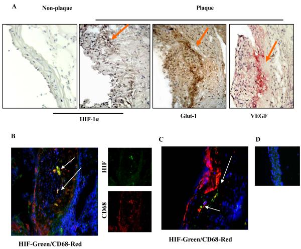
Rationale: Human atherosclerotic plaques contain large numbers of cells deprived of O(2). In murine atherosclerosis, because the plaques are small, it is controversial whether hypoxia can occur.

Objective: To examine if murine plaques contain hypoxic cells, and whether hypoxia regulates changes in cellular lipid metabolism and gene expression in macrophages.

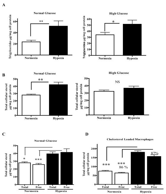
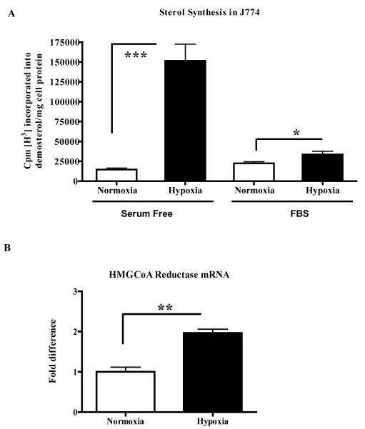
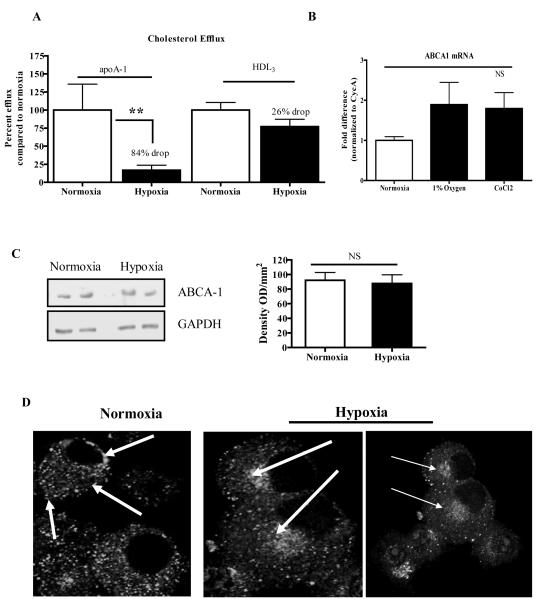
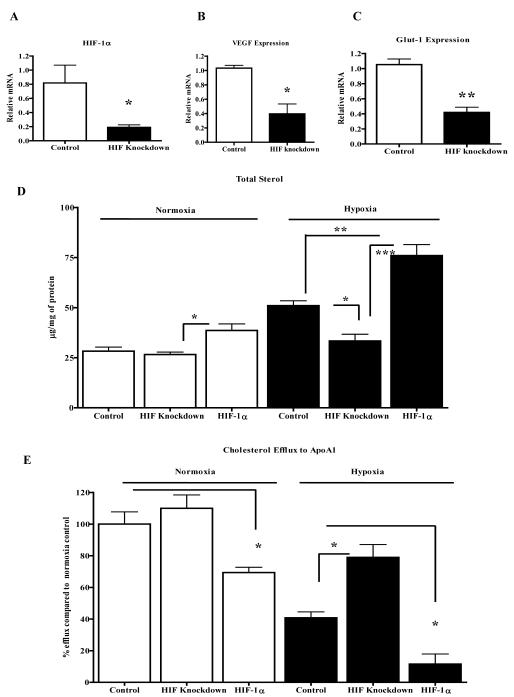
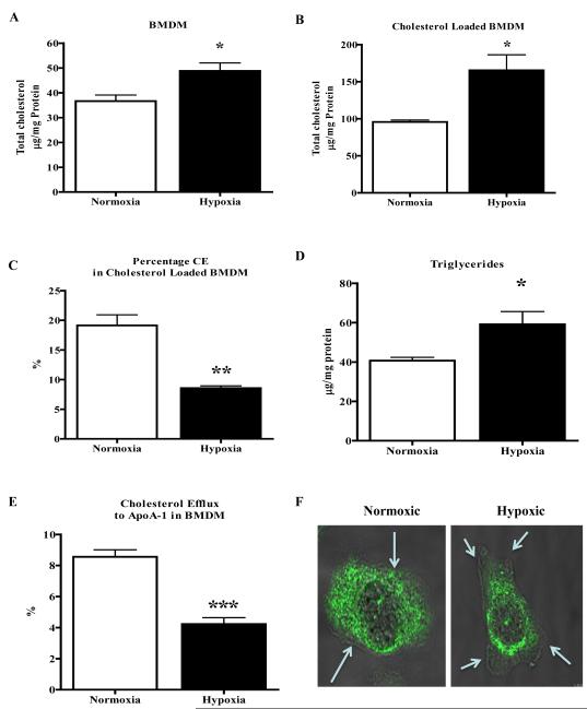
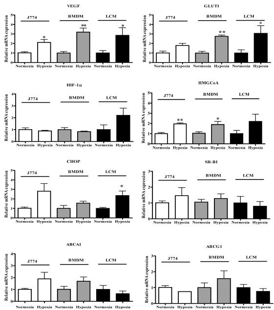
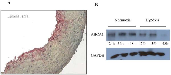
Methods and results: Aortic plaques from apolipoprotein-E-deficient mice were immunopositive for hypoxia-inducible transcription factor (HIF-1α) and some of its downstream targets. Murine J774 macrophages rendered hypoxic demonstrated significant increases in cellular sterol and triglycerides. The increase in sterol content in hypoxic macrophages correlated with elevated 3-hydroxy-3-methyl-glutaryl-CoA (HMG-CoA) reductase activity and mRNA levels. In addition, when macrophages were incubated with cholesterol complexes, hypoxic cells accumulated 120% more cholesterol, predominately in the free form. Cholesterol-efflux assays showed that hypoxia significantly decreased efflux mediated by ATP-binding cassette subfamily A member 1 (ABCA1), whose sub cellular localization was altered in both J774 and primary macrophages. Furthermore, in vivo expression patterns of selected genes from cells in hypoxic regions of murine plaques were similar to those from J774 and primary macrophages incubated in hypoxia. The hypoxia-induced accumulation of sterol and decreased cholesterol efflux was substantially reversed in vitro by reducing the expression of the hypoxia-inducible transcription factor, HIF-1α.

Conclusion: Hypoxic regions are present in murine plaques. Hypoxic macrophages have increased sterol content due to the induction of sterol synthesis and the suppression of cholesterol efflux, effects that are in part mediated by HIF-1α.


Figures

Figure 1
Figure 1. HIF-1α and its target genes are expressed in mouse atherosclerotic plaques
ApoE-/-mice were fed the Western diet for 16 weeks. Aortic roots and arches were harvested and frozen sections were prepared. A, Differential immunohistochemical staining for HIF-1(plaque and healthy vasculature) and two of its well known downstream targets, Glut-1 and VEGF. Staining intensity begins at approximately 100 microns from the vessel lumen; B-C, To examine macrophages in hypoxic areas, we performed double immunofluoresence studies using antibodies to HIF-1α (green) and CD68, a macrophage marker (red). Nuclei were stained with DAPI (blue). Two separate examples are shown, with typical results for the individual antibodies in the small images in panel B; D, Double immunofluoresence experiments were also performed on non-diseased areas of the aortic arch. No detectable signals were detected except for DAPI. Arrows highlight areas with representative staining.
Figure 2
Figure 2. J774 macrophage triglyceride and sterol contents are increased by hypoxia under normoglycemic conditions
J774 macrophages were incubated for 24 h under normoxic or hypoxic (1% O2) conditions in either normal or hyperglycemic medium containing 15% FBS (panels A-C) or serum free (panel D). Lipid assays were performed and shown are the effects of hypoxia on cellular levels of: A, Triglycerides; B, Total sterol (desmosterol+cholesterol); C, Total and free sterol in non-cholesterol-loaded cells. Note that the assays for total and free sterol are independently performed, so that minor assay variations may appear as small differences, such as those in the hypoxic cells; D, Same as panel C, but in cholesterol-loaded cells. The majority of total sterol is in the free form (~86-87%) in both conditions, despite the massive increase in sterol content in hypoxia. All panels: These experiments were performed 3 times in triplicate. Statistical significance is * for p<0.05, ** for p<0.01, and *** p<0.001. NS- non significant.
Figure 3
Figure 3. Hypoxia increases sterol synthesis and HMGCoA reductase mRNA in J774 macrophages
J774 macrophages were incubated for 24h under normoxic or hypoxic (1% O2) conditions in either serum-free or rich (15% FBS) medium. A, Newly synthesized sterol was assessed by incorporation of radioactive acetate into desmosterol under the conditions indicated; B, HMGCoA reductase mRNA was assessed by qRT-PCR from cells in serum-containing medium. This is a representative of 3 experiments performed in triplicate. Statistical significance is indicated by *p<0.05, ** p<0.01, and *** p<0.001.
Figure 4
Figure 4. Cholesterol efflux from J774 macrophages is decreased by hypoxia concurrent with the sub-cellular redistribution of ABCA-1
A, J774 macrophages were incubated for 24 h under normoxic and hypoxic (1% O2) conditions, and efflux of radio-labeled free cholesterol to lipid-poor apoAI (an ABCA-1 acceptor) or HDL3 (an ABCG1 or SR-BI acceptor) was measured (Methods); B, ABCA-1 mRNA abundance was measured under hypoxic or hypoxia-mimicking (CoCl2) conditions; C, ABCA-1 protein levels were detected by western blot (representative sample on left panel) and quantified by densitometry (right panel); D, Confocal microscopic images of indirect immunofluorescence of ABCA-1. The arrows highlight that the distribution of ABCA-1 shifted from the plasma membrane and in the cytoplasm under normoxic conditions to a juxtanuclear location in hypoxia. For the graphs, statistical significance is represented * for p<0.05 and ns for non significance. Efflux experiments were performed 3 times in triplicate and ABCA-1 expression levels were performed twice in duplicate.
Figure 5
Figure 5. HIF-1α plays a direct role in macrophage sterol metabolism A-C
J774 cells were transfected to stably express HIF-1α shRNA or scrambled shRNA (“Control”) and were incubated under hypoxic conditions (1% O2). Shown are the resulting levels of the indicated mRNA species; D, J774 cells were stably transfected either with control plasmid (pBabe-puro), scrambled shRNA (“Control”), HIF-1α shRNA (“HIF Knockdown”) or a HIF-1α expression plasmid (“HIF-1α”), and desmosterol+cholesterol (“Total Sterol”) cellular content measured after incubation in normoxic (left) and hypoxic (right) conditions for 24 h; E, J774 cells were treated as in panel D, but now cholesterol efflux to apoAI, which is the acceptor for ABCA-1, was measured. Experiments were done twice in duplicate (A-C) or quadruplicates (D and E) and statistical significance is indicated by * for p<0.05, **p<0.01. The data with plasmid (pBabe-puro), and scrambled shRNA samples were essentially the same and were combined as one control group.
Figure 6
Figure 6. Bone marrow derived macrophages (BMDM) show altered cholesterol metabolism under hypoxic conditions
A-F, BMDM cells from apoE-/- mice were incubated under hypoxic conditions (1% O2) for 24h. Shown are the resulting levels of total cholesterol from cells that were non-loaded (A) or loaded with cholesterol-cyclodextrin complexes (B). C, The percentage of total cholesterol that was cholesteryl ester was decreased in BMDM loaded with cholesterol under hypoxic conditions. D, BMDM also showed elevated triglycerides under hypoxic conditions. E, BMDM cells were used to measure free cholesterol efflux to apoAI, which is the acceptor for ABCA-1, under normoxic and hypoxic conditions. F, BMDM cells rendered hypoxic were stained for ABCA-1 using confocal microscopy and overlaid with phase contrast image using 63X objective. The arrows indicate the plasma membrane boundary. Experiments were done twice in triplicate (A-E) and statistical significance is indicated by * for p<0.05, ** for p<0.01 and ***p<0.001.
Figure 7
Figure 7. Hypoxic regions of atherosclerotic plaques show a gene expression pattern similar to those in macrophages incubated under hypoxic conditions
ApoE-/- mice were fed the Western diet for 16 weeks and aortic root sections were prepared for laser capture microdissection. Samples were isolated from normoxic and hypoxic regions (positive for both VEGF and GLUT-1 staining; on-line Methods). J774 macrophages and BMDM were exposed to hypoxia in vitro for 24 h. RNA was isolated from laser-captured samples and cultured macrophages, and mRNA abundances was measured using qRT-PCR with resulted normalized to Cyclophilin A. Note that the graphs for ABCA-1 and HMGCoA reductase are repeated from Figures 4B and 3B, respectively. Statistical significance is indicated by * for p<0.05 and ** for p<0.01. The in vitro experiments were performed 3 times in triplicate and the LCM samples were isolated from 6 animals.
Figure 8
Figure 8. Decreased expression of ABCA1 protein in deeper regions of plaques and in macrophages maintained under prolonged hypoxia
A, ApoE-/- mice were fed Western diet for 16 weeks and aortic root sections were prepared. Note that by immunostaining, ABCA1 protein was more abundant in the adlumenal plaque; B, Western blot analysis of ABCA1 protein in lysates of J774 macrophages incubated under hypoxic conditions for 24 h or 48 h. The GAPDH levels are shown for each lane. Results are representative of 3 separate experiments.

Similar articles

Cited by

References

    1. Davidson MH. Overview of prevention and treatment of atherosclerosis with lipid-altering therapy for pharmacy directors. Am J Manag Care. 2007;13(Suppl 10):S260–269. - PubMed
    1. Mensah GA, Brown DW. An overview of cardiovascular disease burden in the united states. Health Aff (Millwood) 2007;26:38–48. - PubMed
    1. Levin M, Leppanen O, Evaldsson M, Wiklund O, Bondjers G, Bjornheden T. Mapping of atp, glucose, glycogen, and lactate concentrations within the arterial wall. Arterioscler Thromb Vasc Biol. 2003;23:1801–1807. - PubMed
    1. Bjornheden T, Levin M, Evaldsson M, Wiklund O. Evidence of hypoxic areas within the arterial wall in vivo. Arterioscler Thromb Vasc Biol. 1999;19:870–876. - PubMed
    1. Leineweber C, Kecklund G, Janszky I, Akerstedt T, Orth-Gomer K. Snoring and progression of coronary artery disease: The stockholm female coronary angiography study. Sleep. 2004;27:1344–1349. - PubMed

Publication types

MeSH terms