. 2012 Jan 1;302(1):G105-15.
doi: 10.1152/ajpgi.00194.2011. Epub 2011 Sep 15.

Spontaneous autoimmune gastritis and hypochlorhydria are manifest in the ileitis-prone SAMP1/YitFcs mice

Affiliations

Spontaneous autoimmune gastritis and hypochlorhydria are manifest in the ileitis-prone SAMP1/YitFcs mice

P B Ernst et al. Am J Physiol Gastrointest Liver Physiol. .

Abstract

SAMP1/YitFcs mice serve as a model of Crohn's disease, and we have used them to assess gastritis. Gastritis was compared in SAMP1/YitFcs, AKR, and C57BL/6 mice by histology, immunohistochemistry, and flow cytometry. Gastric acid secretion was measured in ligated stomachs, while anti-parietal cell antibodies were assayed by immunofluorescence and enzyme-linked immunosorbent spot assay. SAMP1/YitFcs mice display a corpus-dominant, chronic gastritis with multifocal aggregates of mononuclear cells consisting of T and B lymphocytes. Relatively few aggregates were observed elsewhere in the stomach. The infiltrates in the oxyntic mucosa were associated with the loss of parietal cell mass. AKR mice, the founder strain of the SAMP1/YitFcs, also have gastritis, although they do not develop ileitis. Genetic studies using SAMP1/YitFcs-C57BL/6 congenic mice showed that the genetic regions regulating ileitis had comparable effects on gastritis. The majority of the cells in the aggregates expressed the T cell marker CD3 or the B cell marker B220. Adoptive transfer of SAMP1/YitFcs CD4(+) T helper cells, with or without B cells, into immunodeficient recipients induced a pangastritis and duodenitis. SAMP1/YitFcs and AKR mice manifest hypochlorhydria and anti-parietal cell antibodies. These data suggest that common genetic factors controlling gastroenteric disease in SAMP1/YitFcs mice regulate distinct pathogenic mechanisms causing inflammation in separate sites within the digestive tract.


Figures
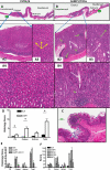
Fig. 1.
Fig. 1.
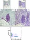
SAMP1/YitFcs mice develop a corpus-dominant gastritis. Gastric tissue was collected from control C57BL/6 mice (A) and SAMP1/YitFcs mice (B), and inflammation was assessed by histology. A: normal, uninflamed gastric mucosa. Higher power (A2) shows a typical uniform population of parietal cells in the corpus; representative parietal cells are indicated by yellow arrows in A3. Gastric mucosa of SAMP1/YitFcs mice is typically thickened (B; scale bars, ∼500 μm in A and B). At higher magnification, mononuclear cell aggregates are apparent in the corpus (green arrows, B2B4); dilated glands containing accumulations of dead cells are indicated by white arrows (B2B4). Images captured at a higher power show intact parietal cells in control mouse (A4) and inflammation with associated loss of parietal cell mass in SAMP1/YitFcs mouse (B4). Aggregates (encircled) were observed in the forestomach of some mice (C; scale bar, ∼100 μm). D: scoring for forestomach (FS), corpus, and antrum from 5 SAMP1/YitFcs (SAMP1) mice and 6 C57BL/6 (BL/6) age-matched mice, representing a sample of >60 mice. Values are means ± SE. *P < 0.05. As several genetic elements regulating the onset of ileitis in SAMP1/YitFcs mice have been identified, gastritis was assessed in these crosses. E: scoring for the forestomach, corpus, and antrum from 10 SAMP1/YitFcs, 9 AKR, and 9 (AKR × SAMP1/YitFcs) F1 mice. F: gastritis scores for 10 C57BL/6 mice and 13 SAMP1/YitFcs mice compared with scores for SAMP1/Yit/Fcs-C57BL/6 congenic mice, including 8 SAMP.B6C9A (B6C9A), 3 SAMP.B6C9BL (B6C9BL), and 3 SAMP.B6CX (B6CX) mice. Values are means ± SE. *P < 0.05 vs. SAMP1.
Fig. 2.
Fig. 2.
Characterization of mononuclear cells within the aggregates. Photomicrographs represent 2 magnifications of sections stained for B220 (A and C) and CD3 (B and D). Number of B220+ cells within these aggregates exceeded the number of CD3+ cells. Images are from 1 mouse and are representative of 5 separate SAMP1/YitFcs mice. E: flow cytometric analysis of mononuclear cells isolated from the gastric corpus lamina propria and stained for B220 and CD3. Results from representative experiment show that almost all the freshly isolated gastric mononuclear cells expressed CD3 or B220, while a significant subset expressed both markers. PE, phycoerythrin.
Fig. 3.
Fig. 3.
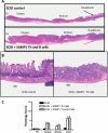
Adoptive transfer of CD4+ T helper (Th) cells induces a pangastritis and duodenitis distinct from the spontaneous disease in SAMP1/YitFcs mice. To investigate the role of Th cells in the pathogenesis of gastritis, severe combined immunodeficiency (SCID) recipient mice were given 5 × 10⁵ CD4+ Th cells with or without an equal number of B cells, both prepared from the mesenteric lymph node of SAMP1/YitFcs mice. After 6 wk, tissues were collected. A: control SCID tissue had little inflammation and a completely healthy duodenum (black arrow). SCID recipients given Th cells and B cells from SAMP1/YitFcs mice (same magnification as SCID control) had a slightly thickened corpus and evidence of a generalized pangastritis, with more obvious lesions in the antrum in the recipients than the donor SAMP1/YitFcs mice. In addition, their duodenal architecture was virtually obliterated (red arrow and higher power in B; scale bar, ∼100 μm). Parietal cells are obvious and intact in these recipients. C: histological scoring. Data are from 2 SCID and 4 reconstituted mice per group.
Fig. 4.
Fig. 4.
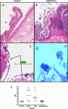
SAMP1/YitFcs mice have evidence of bacterial overgrowth and achlorhydria. Hyperkeratosis was observed in the forestomach in ∼50% of the SAMP1/YitFcs mice (B and C) but was not evident in the C57BL/6 mice (A). In some cases, a marked accumulation of rod-shaped bacteria was evident (D). These findings suggest that there may be bacterial overgrowth due to altered gastric acid production. Examination of the gastric pH over 3 h in unstimulated, ligated stomachs revealed abnormal gastric acid production that reached pH 7 in some cases in SAMP1/YitFcs and AKR mice, suggestive of achlorhydria (E). (AKR × SAMP1/YitFcs) F1 mice, which lacked gastritis, had essentially normal gastric acid production. Data are from 6–9 mice per group. Values are median scores. *P < 0.05 vs. C57BL/6 mice (Wilcoxon's rank sum test).
Fig. 5.
Fig. 5.
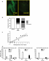
SAMP1/YitFcs and AKR mice have an autoimmune gastritis and produce anti-parietal cell antibodies. Given the effect on gastric pH, SAMP1/YitFcs (n = 6), AKR (n = 3), and (AKR × SAMP1/YitFcs) F1 mice (n = 5) were assayed for anti-parietal cell antibodies and compared with C57BL/6 mice (n = 6). A: representative immunofluorescence image in which serum from SAMP1/YitFcs mice (left) binds selectively to the parietal cells, in contrast to the sample from C57BL/6 mice (right). B: percentage of each strain with titer scores of 0 (negative), 1 (+), 2 (++), or 3 (+++). C: analysis of the inflammation score in the corpus of all 20 samples and the titers of anti-parietal cell antibodies showed a significant correlation between the 2 variables (r = 0.9274, P < 0.05). D: number of nonspecific (IgM, IgG, or IgA) and antigen-specific antibody-producing cells in gastric mononuclear cells (gMNC), spleen (SPL), and bone marrow (BM) isolated from SAMP1/YitFcs mice detected by enzyme-linked immunosorbent spot assay. Values are means ± SE for 3 observations.

Similar articles

References

    1. Alam MS, Kurtz CC, Rowlett RM, Reuter BK, Wiznerowicz E, Das S, Linden J, Crowe SE, Ernst PB. CD73 is expressed by human regulatory T helper cells and suppresses proinflammatory cytokine production and Helicobacter felis-induced gastritis in mice. J Infect Dis 199: 494–504, 2009 - PMC - PubMed
    1. Alam MS, Kurtz CC, Wilson JM, Burnette BR, Wiznerowicz EB, Ross WG, Rieger JM, Figler RA, Linden J, Crowe SE, Ernst PB. A2A adenosine receptor (AR) activation inhibits pro-inflammatory cytokine production by human CD4+ helper T cells and regulates Helicobacter-induced gastritis and bacterial persistence. Mucosal Immunol 2: 232–242, 2009 - PMC - PubMed
    1. Bamford KB, Fan XJ, Crowe SE, Leary JF, Gourley WK, Luthra GK, Brooks EG, Graham DY, Reyes VE, Ernst PB. Lymphocytes in the human gastric mucosa during Helicobacter pylori have a T helper cell 1 phenotype. Gastroenterology 114: 482–492, 1998 - PubMed
    1. Bamias G, Martin C, Mishina M, Ross WG, Rivera-Nieves J, Marini M, Cominelli F. Proinflammatory effects of TH2 cytokines in a murine model of chronic small intestinal inflammation. Gastroenterology 128: 654–666, 2005 - PubMed
    1. Bamias G, Okazawa A, Rivera-Nieves J, Arseneau KO, De La Rue SA, Pizarro TT, Cominelli F. Commensal bacteria exacerbate intestinal inflammation but are not essential for the development of murine ileitis. J Immunol 178: 1809–1818, 2007 - PubMed

Publication types

MeSH terms