. 2012 Jan 1;302(1):G44-54.
doi: 10.1152/ajpgi.00413.2010. Epub 2011 Sep 15.

Hydrogen sulfide preconditioning or neutrophil depletion attenuates ischemia-reperfusion-induced mitochondrial dysfunction in rat small intestine

Affiliations

Hydrogen sulfide preconditioning or neutrophil depletion attenuates ischemia-reperfusion-induced mitochondrial dysfunction in rat small intestine

Yajun Liu et al. Am J Physiol Gastrointest Liver Physiol. .

Abstract

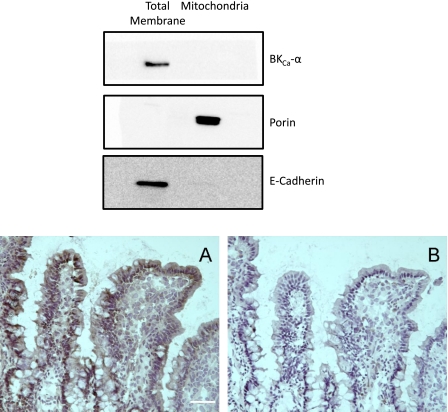
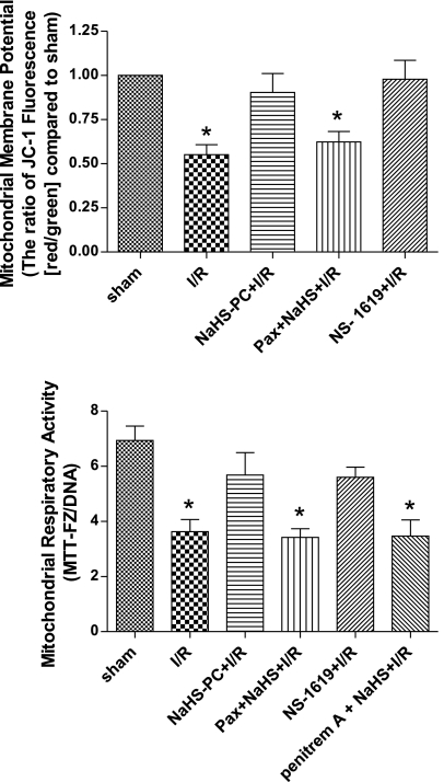
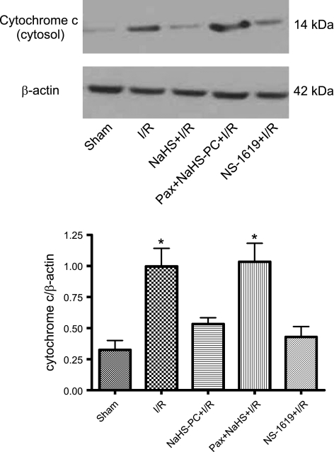
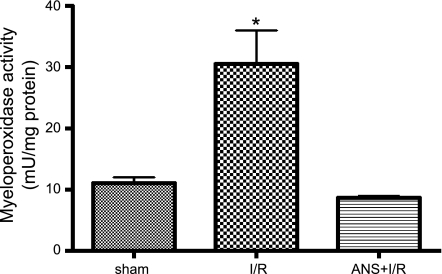
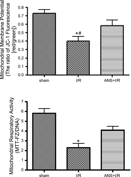
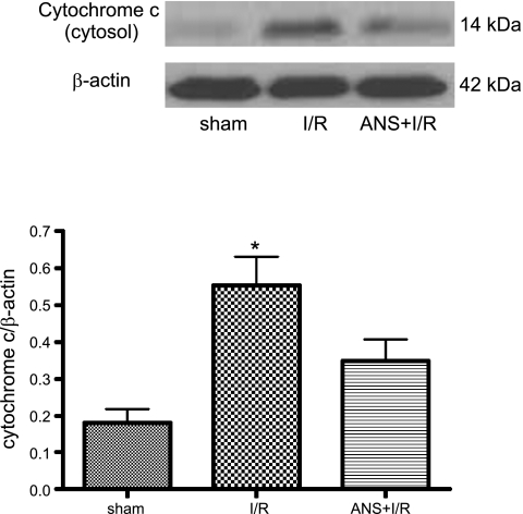
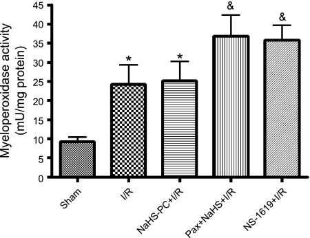
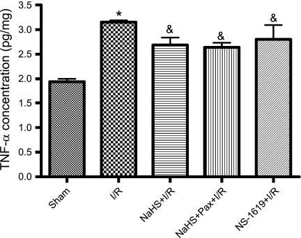
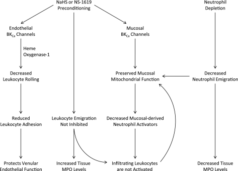
The objectives of this study were to determine whether neutrophil depletion with anti-neutrophil serum (ANS) or preconditioning with the hydrogen sulfide (H(2)S) donor NaHS (NaHS-PC) 24 h prior to ischemia-reperfusion (I/R) would prevent postischemic mitochondrial dysfunction in rat intestinal mucosa and, if so, whether calcium-activated, large conductance potassium (BK(Ca)) channels were involved in this protective effect. I/R was induced by 45-min occlusion of the superior mesenteric artery followed by 60-min reperfusion in rats preconditioned with NaHS (NaHS-PC) or a BK(Ca) channel activator (NS-1619-PC) 24 h earlier or treated with ANS. Mitochondrial function was assessed by measuring mitochondrial membrane potential, mitochondrial dehydrogenase function, and cytochrome c release. Mucosal myeloperoxidase (MPO) and TNF-α levels were also determined, as measures of postischemic inflammation. BK(Ca) expression in intestinal mucosa was detected by immunohistochemistry and Western blotting. I/R induced mitochondrial dysfunction and increased tissue MPO and TNF-α levels. Although mitochondrial dysfunction was attenuated by NaHS-PC or NS-1619-PC, the postischemic increases in mucosal MPO and TNF-α levels were not. The protective effect of NaHS-PC or NS-1619-PC on postischemic mitochondrial function was abolished by coincident treatment with BK(Ca) channel inhibitors. ANS prevented the I/R-induced increase in tissue MPO levels and reversed mitochondrial dysfunction. These data indicate that neutrophils play an essential role in I/R-induced mucosal mitochondrial dysfunction. In addition, NaHS-PC prevents postischemic mitochondrial dysfunction (but not inflammation) by a BK(Ca) channel-dependent mechanism.


Figures

Fig. 1.
Fig. 1.
Expression of calcium-activated, large conductance potassium (BKCa) channel in rat intestinal enterocytes. Top: Western blot of total membrane and crude mitochondrial fractions from isolated mouse ileal enterocytes (see methods), each probed for the α-subunit of BKCa channel, mitochondrial porin (VDAC, outer membrane marker for mitochondria), and E-cadherin (plasma membrane protein used as marker for the total membrane fraction). Bottom, A: immunostaining of BKCa α-subunit protein in enterocytes of rat ileal mucosa. Bottom, B: negative controls were performed by using BKCa-α primary antibody that had been preabsorbed by use of a control peptide. Scale bar = 20 μm.
Fig. 2.
Fig. 2.
Reductions in mitochondrial membrane potential (top) and respiratory activity (bottom) induced by ischemia-reperfusion (I/R) are prevented by preconditioning with the hydrogen sulfide donor NaHS (NaHS-PC+I/R) or the BKCa channel activator NS-1619 (NS-1619+I/R) 24 h earlier. The effects of NaHS-PC to preserve postischemic mitochondrial function are prevented by coincident administration of the BKCa channel inhibitor paxilline (Pax+NaHS-PC+I/R) or penitrem A (penitrem A+NaHS-PC+I/R) with NaHS 24 h prior to I/R. Data are means ± SE for 6 rats per treatment group. *P < 0.05 compared with sham, NaHS-PC+I/R and NS-1619+I/R groups.
Fig. 3.
Fig. 3.
I/R induced release of cytochrome c into the cytosol of mucosal epithelial cells relative to sham (nonischemic) intestine. This disruption in postischemic mitochondrial membrane integrity was prevented by preconditioning with either the hydrogen sulfide (H2S) donor NaHS (NaHS-PC+I/R) or the BKCa channel activator NS-1619 (NS-1619+I/R) 24 h prior to induction of I/R. Coincident treatment with paxilline, a BKCa channel inhibitor, with NaHS 24 h prior to I/R (Pax+NaHS-PC+I/R) abrogated the protective actions of this H2S donor on postischemic mitochondrial membrane integrity. Top: representative Western blot showing comparison of signals from cytochrome c vs. β-actin. Bottom: densitometric analysis of full data set from 6 rats per treatment group. *P < 0.05 compared with sham, NaHS-PC+I/R, and NS-1619+I/R groups.
Fig. 4.
Fig. 4.
Increase in ileal mucosal myeloperoxidase (MPO) activity induced by I/R relative to sham (nonischemic) controls is prevented by neutrophil depletion by use of anti-neutrophil serum (ANS). Data are means ± SE for 6 rats per treatment group. *P < 0.001 compared with sham and ANS+I/R groups.
Fig. 5.
Fig. 5.
Neutrophil depletion with ANS prevents the reductions in mitochondrial membrane potential (top) and respiratory activity (bottom) induced by I/R relative to sham (nonischemic) controls. Data represents means ± SE for 6 rats per group. *P < 0.01 compared with sham group, #P < 0.05 compared with ANS+I/R group. JC-1, 5,5′,6,6′-tetrachloro-1,1′,3,3′-tetraethyl-imidacarbocyanine iodide; MTT-FZ, formazan derivative of 3-(4,5-dimethylthiazol-2-yl)-2,5 diphenyltetrazolium bromide.
Fig. 6.
Fig. 6.
ANS+I/R treatment prevented the postischemic release of cytochrome c into enterocyte cytosol induced by I/R compared with sham (nonischemic) preparations. Top: representative Western blot from full data set. Bottom: densitometric analysis of aggregate data. Data are means ± SE from 6 rats per group. *P < 0.05 compared with sham and ANS+I/R groups.
Fig. 7.
Fig. 7.
I/R induced an increase in intestinal MPO activity relative to sham (nonischemic) controls, an effect that was not attenuated by preconditioning with the H2S donor NaHS (NaHS-PC+I/R) or the BKCa activator NS-1619 (NS-1619+I/R) 24 h prior to I/R. Coincident administration of the BKCa inhibitor paxilline with NaHS 24 h prior to I/R (Pax+NaHS+I/R) was associated with mucosal MPO levels that were significantly elevated relative to sham but were not statistically different from I/R. Data are means ± SE for 6 rats per group. *P < 0.05 compared with sham. &P < 0.001 compared with sham.
Fig. 8.
Fig. 8.
Ileal mucosal tumor necrosis factor-α (TNF-α) levels were increased following I/R. This postischemic increase in mucosal TNF-α was not affected by preconditioning with the H2S donor NaHS (NaHS-PC+I/R) or the BKCa channel activator NS-1619 (NS-1619+I/R) nor by coincident administration of the BKCa channel inhibitor paxilline with NaHS (Pax+NaHS+I/R) 24 h prior to I/R. Data are means ± SE for 6 rats per group. *P < 0.001 compared with sham, &P < 0.05 compared with sham.
Fig. 9.
Fig. 9.
Summary diagram illustrating the role of endothelial and mucosal BKCa channels as mediators of the effects of antecedent treatment with NaHS or NS-1619 (NaHS or NS-1619 Preconditioning) to limit I/R-induced leukocyte rolling and adhesion and preserve mucosal mitochondrial function. Neutrophil depletion with ANS prior to the onset of I/R also preserved mucosal mitochondrial function. Since I/R-induced neutrophil infiltration (assessed using tissue MPO activity as a marker) was prevented by ANS treatment, but not by administration of NaHS or NS-1619, our results suggest that infiltrating neutrophils may not be activated to produce collateral injury in the postischemic intestine of animals preconditioned with NaHS or NS-1619. Although the postischemic increase in TNF-α was not attenuated by NaHS or NS-1619 preconditioning, multiple chemotactic agents are released and act in concert to promote neutrophil in the interstitial space after intestinal I/R. It is possible that release of at least some of these neutrophil activators may be abrogated by preconditioning, thereby limiting activation of extravasated leukocytes.

Similar articles

Cited by

References

    1. Boehning D, Snyder S. Novel neural modulators. Annu Rev Neurosci 26: 105–131, 2003 - PubMed
    1. Bolli R. Preconditioning: a paradigm shift in the biology of myocardial ischemia. Am J Physiol Heart Circ Physiol 292: H19–H27, 2007 - PMC - PubMed
    1. Cho J, Won K, Wu D, Soong Y, Liu S, Szeto HH, Hong MK. Potent mitochondria-targeted peptides reduce myocardial infarction in rats. Coron Artery Dis 18: 215–220, 2007 - PubMed
    1. Cooper D, Russell J, Chitman KD, Williams MC, Wolf RE, Granger DN. Leukocyte dependence of platelet adhesion in postcapillary venules. Am J Physiol Heart Circ Physiol 286: H1895–H1900, 2004 - PubMed
    1. Douglas RM, Lai JC, Bian S, Cummins L, Moczydlowski E, Haddad GG. The calcium-sensitive large-conductance potassium channel (BK/MAXI K) is present in the inner mitochondrial membrane of rat brain. Neuroscience 139: 1249–1261, 2006 - PubMed

Publication types

MeSH terms