. 2011 Oct;26(5):423-33.
doi: 10.1177/0748730411416341.

Disruption of circadian rhythms accelerates development of diabetes through pancreatic beta-cell loss and dysfunction

Affiliations

Disruption of circadian rhythms accelerates development of diabetes through pancreatic beta-cell loss and dysfunction

John E Gale et al. J Biol Rhythms. 2011 Oct.

Abstract

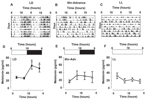
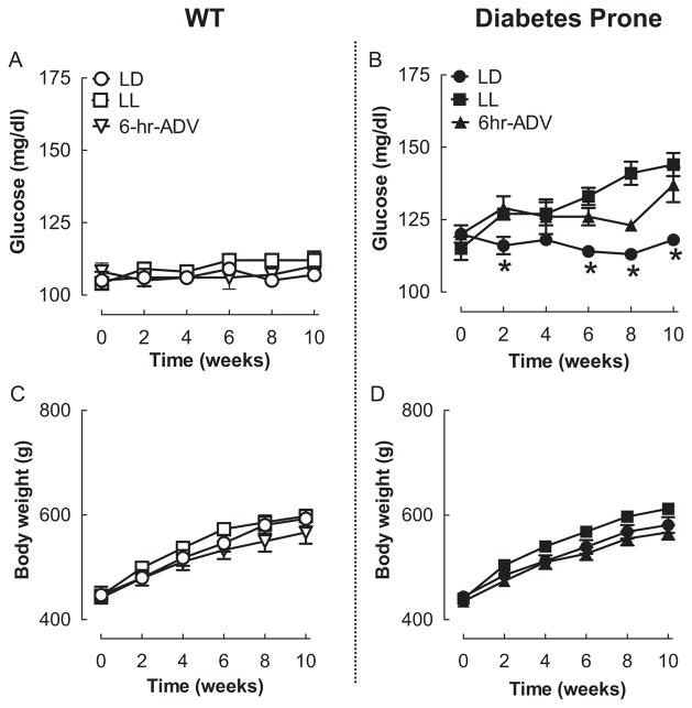
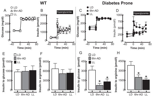
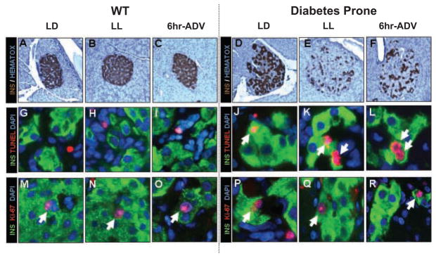
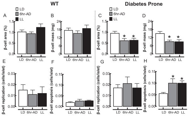
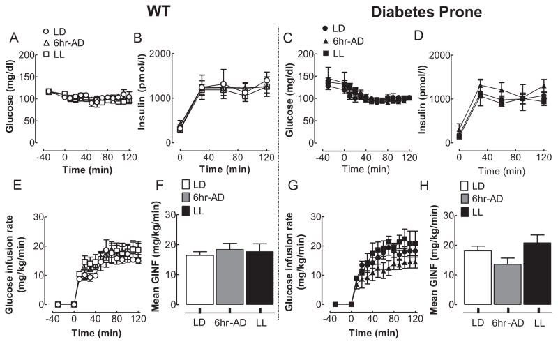
Type 2 diabetes mellitus (T2DM) is complex metabolic disease that arises as a consequence of interactions between genetic predisposition and environmental triggers. One recently described environmental trigger associated with development of T2DM is disturbance of circadian rhythms due to shift work, sleep loss, or nocturnal lifestyle. However, the underlying mechanisms behind this association are largely unknown. To address this, the authors examined the metabolic and physiological consequences of experimentally controlled circadian rhythm disruption in wild-type (WT) Sprague Dawley and diabetes-prone human islet amyloid polypeptide transgenic (HIP) rats: a validated model of T2DM. WT and HIP rats at 3 months of age were exposed to 10 weeks of either a normal light regimen (LD: 12:12-h light/dark) or experimental disruption in the light-dark cycle produced by either (1) 6-h advance of the light cycle every 3 days or (2) constant light protocol. Subsequently, blood glucose control, beta-cell function, beta-cell mass, turnover, and insulin sensitivity were examined. In WT rats, 10 weeks of experimental disruption of circadian rhythms failed to significantly alter fasting blood glucose levels, glucose-stimulated insulin secretion, beta-cell mass/turnover, or insulin sensitivity. In contrast, experimental disruption of circadian rhythms in diabetes-prone HIP rats led to accelerated development of diabetes. The mechanism subserving early-onset diabetes was due to accelerated loss of beta-cell function and loss of beta-cell mass attributed to increases in beta-cell apoptosis. Disruption of circadian rhythms may increase the risk of T2DM by accelerating the loss of beta-cell function and mass characteristic in T2DM.


Conflict of interest statement

CONFLICT OF INTEREST STATEMENT

The author(s) have no potential conflicts of interest with respect to the research, authorship, and/or publication of this article.

Figures

Figure 1
Figure 1
Impact of LL and 6-h advances on diurnal rhythms of activity and plasma melatonin in rats. (A-C) Representative double-plotted actograms of rats exposed to either control light (A) regimen (12:12 h, LD; n = 5) or experimental changes in the LD cycle produced by (B) 6-h advance of the light cycle every 3 days for 10 weeks (6-h ADV; n = 5) or by 24-h constant light (LL; n = 5). (D-F) Diurnal levels of plasma melatonin in WT and HIP rats following 10-week exposure to LD (D), LL (E), or 6-h ADV (F). Data are expressed as mean ± SEM, *p < 0.05 statistical significance.
Figure 2
Figure 2
Effects of 10-week circadian rhythm disruption on fasting glucose and body weight. Influence of environmental circadian rhythm disruption on fasting glucose (A, B) and body weight (C, D) in WT (left panels) and diabetes-prone HIP (right panels) rats exposed to 10 weeks of LD (open and closed circles), LL (open and filled squares), or 6-h advance (open and filled triangles) regimens. Data are expressed as mean ± SEM. *p < 0.05 statistical significance for changes in plasma glucose in HIP LD light vs. HIP 6-h ADV and HIP LL.
Figure 3
Figure 3
Effects of 10-week circadian rhythm disruption on pancreatic beta-cell function in WT and HIP rats. Mean plasma glucose (A, C) and insulin (B, D) concentrations during the hyperglycemic clamp in WT (left panels) and diabetes-prone HIP rats (right panels) following 10-week exposure to LD (open and closed circles), LL (open and filled squares), or 6-h advance (open and filled triangles) regimen. Mean insulin response to glucose (E, G) and arginine (F, H) challenge during the hyperglycemic clamp in WT (left panel) and diabetes-prone HIP (right panels) rats following 10-week exposure to LD (open and closed circles), LL (open and filled squares), or 6-h advances (open and filled triangles) regimen. Data are expressed as mean ± SEM. *p < 0.05 statistical significance for insulin response to glucose and arginine vs. LD HIP.
Figure 4
Figure 4
Effects of 10-week circadian rhythm disruption on pancreatic beta-cell mass and turnover. (A-F) Representative examples of pancreatic islets stained by immunohistochemistry for insulin (brown) and nuclear stain hematoxylin (blue) imaged at 20x in WT (left panels) and diabetes-prone HIP rats (right panels) following 10-week exposure to LD (A, D), LL (B, E), and 6-h advance (C, F) regimens. (G–L) Representative examples of pancreatic islets stained by immunofluorescence for insulin (green), marker of cell apoptosis TUNEL (red), and nuclear stain DAPI (blue) and (M–R) representative examples of pancreatic islets stained by immunofluorescence for insulin (green), marker of cell replication Ki-67 (red), and nuclear stain DAPI (blue) imaged at 60x in WT (left panels) and diabetes-prone HIP rats (right panels) following 10-week exposure to LD (M, P), LL (N, Q), and 6-h advances (O, R).
Figure 5
Figure 5
Effects of 10-week circadian rhythm disruption on pancreatic beta-cell mass, beta-cell apoptosis, and replication. Mean beta-cell fractional area (A, C) and beta-cell mass (B, D) in WT (left panels) and diabetes-prone HIP rats (right panels) following 10-week exposure to LD (open bars), LL (black bars), and 6-h advances (gray bars). Mean beta-cell replication (E, G) and beta-cell apoptosis (F, H) in WT (left panels) and diabetes-prone HIP rats (right panels) following 10-week exposure to LD (open bars), LL (black bars), and 6-h advances (gray bars). Data are expressed as mean ± SEM. *p < 0.05 statistical significance for beta-cell area, beta-cell mass, and beta-cell apoptosis vs. LD HIP rats.
Figure 6
Figure 6
Effects of 10-week circadian rhythm disruption on insulin sensitivity in WT and diabetes-prone HIP rats. Mean glucose (A, C) and plasma insulin (B, D) concentrations during the hyperinsulinemic-euglycemic clamp (0–120 min) in WT (left panels) and diabetes-prone HIP rats (right panels) following 10-week exposure to LD (open and closed circles), LL (open and filled squares), or 6-h advances (open and filled triangles). Corresponding (E, G) and cumulative (F, H) glucose infusion rates during the hyperinsulinemic-clamp in WT (left panels) and diabetes-prone HIP rats (right panels) following 10-week exposure to LD (open and closed circles), LL (open and filled squares), or 6-h advances (open and filled triangles). Data are expressed as mean ± SE.

Similar articles

Cited by

References

    1. Bass J, Takahashi JS. Circadian integration of metabolism and energetics. Science. 2010;330:1349–1354. - PMC - PubMed
    1. Bergman RN, Phillips LS, Cobelli C. Physiologic evaluation of factors controlling glucose tolerance in man: Measurement of insulin sensitivity and beta-cell glucose sensitivity from the response to intravenous glucose. J Clin Invest. 1981;68:1456–1467. - PMC - PubMed
    1. Bouatia-Naji N, Bonnefond A, Cavalcanti-Proenca C, Sparso T, Holmkvist J, Marchand M, Delplanque J, Lobbens S, Rocheleau G, Durand E, et al. A variant near MTNR1B is associated with increased fasting plasma glucose levels and type 2 diabetes risk. Nat Genet. 2009;41:89–94. - PubMed
    1. Butler AE, Jang J, Gurlo T, Carty MD, Soeller WC, Butler PC. Diabetes due to a progressive defect in beta-cell mass in rats transgenic for human islet amyloid polypeptide (HIP rat): A new model for type 2 diabetes. Diabetes. 2004;53:1509–1516. - PubMed
    1. Butler AE, Janson J, Bonner-Weir S, Ritzel R, Rizza RA, Butler PC. Beta-cell deficit and increased beta-cell apoptosis in humans with type 2 diabetes. Diabetes. 2003;52:102–110. - PubMed

Publication types