

Review
doi: 10.4161/viru.2.5.17724.
Epub 2011 Sep 1.
Staphylococcus aureus biofilms: properties, regulation, and roles in human disease
Affiliations
- PMID: 21921685
- PMCID: PMC3322633
- DOI: 10.4161/viru.2.5.17724
Item in Clipboard
Review
Staphylococcus aureus biofilms: properties, regulation, and roles in human disease
Virulence.
2011 Sep-Oct.
Abstract
Increasing attention has been focused on understanding bacterial biofilms and this growth modality's relation to human disease. In this review we explore the genetic regulation and molecular components involved in biofilm formation and maturation in the context of the Gram-positive cocci, Staphylococcus aureus. In addition, we discuss diseases and host immune responses, along with current therapies associated with S. aureus biofilm infections and prevention strategies.
Figures

IgGs against recombinant forms of cell wall-associated biofilm proteins bind to intact S. aureus biofilms. IgG against each selected candidate protein was applied followed by the secondary goat anti-rabbit F(ab′)2 antibody (upper right in A–D). After a washing step, SYTO 9 was applied to stain all bacterial cells (upper left in A–D). Biofilms were probed with (A) anti-SA0486 (autolysin) IgG and secondary antibody, (B) anti-SA0486 (lipoprotein) IgG and secondary antibody, (C) anti-SA0688 (lipoprotein) IgG and secondary antibody and (D) anti-SA0037 (conserved hypothetical) IgG and secondary antibody. The merged image is shown in the lower part in (A–D). The base of the glass is located at the bottom of each image, and each image is a cross-sectional view of the biofilm from the base into the lumen.

SEM micrograph from an implant recovered at five days showing massive numbers of cocci partially occluded by dehydrated material. Bar = 5 µm.

TEM micrograph of a cross-section through a biofilm recovered from an indwelling medical device implanted in the peritoneum of a rat for five days. Bacteria are apparent on the surface of the biofilm and interspersed with host material and lysed bacteria within the biofilm. Bar = 5 µm.

TEM micrograph depicts bacteria within a medical implant biofilm. The bacteria are associated with large amounts of host extracellular matrix materials and lysed bacteria. Fibrils are apparent radiating from the bacterial cell surface in a clear “halo” surrounding the bacteria (arrow). Bar = 1 µm.

Flowchart of regulatory factors involved in S. aureus biofilm formation, maintenance and detachment. (A) PIA-dependent biofilm formation—expression of the icaADBC gene cluster results in PIA expression and biofilm formation. Expression of icaADBC can be suppressed by production of tcaR and icaR, resulting in downregulation of PIA and thus biofilm formation., In the case of the icaR gene, expression can be up or downregulated by the proteins Spx and Rbf, respectively., Consequently, Spx induction of icaR expression results in downregulation of icaADBC expression, PIA production and biofilm formation. Conversely, Rbf inhibits icaR expression leading to upregulation of icaADBC expression, PIA production and biofilm formation. Additionally, anaerobic conditions induce production of SrrAB, causing expression of the icaADBC gene cluster, PIA production and biofilm formation. PIA-independent biofilm formation*—in ica-deletion mutants, PIA-independent biofilm formation can be mediated through cell wall-associated protein cell-to-cell adhesion (MRSA-specific). Examples of proteins arbitrating biofilm formation include SpA, FnBPs and Bap.,,
icaADBC, intercellular adhesion biosynthetic genes; PIA, polysaccharide intercellular antigen; tcaR, transcriptional regulator of the teicoplanin-associated locus; icaR, intercellular adhesion regulatory gene; Spx, global regulator of stress response genes; Rbf, protein regulator of biofilm formation; SrrAB, staphylococcal respiratory response regulator; SpA, S. aureus protein A; FnBPs, fibronectin-binding proteins; Bap, biofilm-associated protein. (B) eDNA and biofilm formation—eDNA leads to enhanced biofilm formation.– DNase treatment degrades eDNA and inhibits eDNA-mediated biofilm formation.,, DNA release is arbitrated through cell lysis and controlled by lrg and cidA gene expression., Upregulation of the lrg gene results in inhibition of cellular lysis, DNA release and biofilm formation. Conversely, cidA gene expression enhances cellular lysis, DNA release and biofilm formation. eDNA, extracellular DNA; lrg, regulator of murein hydrolase and cell death; cidA, regulator of murein hydrolase and cell death. (C) agr/sarA/sigB regulation—expression of the sarA gene results in downregulation of proteases and the thermostable nuclease, allowing for development of an immature biofilm. Expression of sigB similarly downregulates protease production, but additionally promotes expression of adherence factors that aid in initial biofilm formation.– The immature biofilm increases in cell density until a mature biofilm develops. At this stage, the density of AIPs throughout the bacterial community reaches a quorum sensing threshold and induces expression of the agr gene. Induction of agr results in upregulation of detergent-like peptide, protease and thermostable nuclease expression; leading to release of bacterial cells from the mature biofilm, termed seeding dispersal.,–
sarA, staphylococcal accessory regulator; sigB, sigmaB; AIP, auto-inducing peptides; agr, accessory gene regulator.
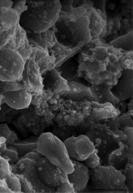
SEM micrograph demonstrating cocci adherent to a bone sequestra. Collapsed glycocalyx material is apparent associated with the bacterial biofilm (arrow). Bar = 5 µm.

(A) Nasal colonization confers Staphylococcus aureus a reservoir for dissemination onto skin and various other host epithelial surfaces. Damage to the epithelia can occur from extraneous sources. (B) Damage allows S. aureus to breach the epithelial layer and bind to host matrix via surface expressed colonization factors. Map, MHC class II analog protein; Cna, collagen-binding adhesin; FnbB, fibronectin binding protein B; FnbA, fibronectin binding protein A; Fib, fibrinogen binding protein; FbpA, fibronectin binding protein; CflA, clumping factor A; EbpS, elastin binding protein. (C) Initial attachment and cell division produces an early S. aureus biofilm. The quorum sensing compound of S. aureus, termed auto-inducing peptide (AIP), is secreted and taken up by the microcolony. SarA expression is upregulated, leading to production of virulence and immunoavoidance factors (CHIPS, chemotaxis inhibitory protein of staphylococci; Eap, extracellular adherence protein; SCIN, staphylococcal complement inhibitor). Additionally, AIP has reached quorum threshold levels, resulting in agr activation and a downregulation of adhesins and an upregulation of virulence factor expression that cause damage to the host and evade the immune response. The nascent S. aureus biofilm avoids immunological destruction by initiating leukocyte apoptosis and/or developing a protective environment via inflammation and tissue damage. (D) Mature S. aureus biofilm is encapsulated by polysaccharide intercellular antigen (PIA), protein and extracellular DNA (eDNA). AIP has reached quorum threshold levels, resulting in agr gene expression. Subsequently, protease and detergent-like peptides are secreted into the biofilm and promote seeding dispersal. Large biofilm aggregates detach forming flocs and planktonic bacterial cells migrate from the mature biofilm into the circulatory system, where adherence to distal tissue and formation of a nascent biofilm can occur to repeat the cycle.
References
-
- Kristian SA, Golda T, Ferracin F, Cramton SE, Neumeister B, Peschel A, et al. The ability of biofilm formation does not influence virulence of Staphylococcus aureus and host response in a mouse tissue cage infection model. Microb Pathog. 2004;36:237–245. doi: 10.1016/j.mic-path.2003.12.004. - DOI - PubMed
-
- Brady RA, Leid JG, Kofonow J, Costerton JW, Shirtliff ME. Immunoglobulins to surface-associated biofilm immunogens provide a novel means of visualization of methicillin-resistant Staphylococcus aureus biofilms. Appl Environ Microbiol. 2007;73:6612–6619. doi: 10.1128/AEM.00855-07. - DOI - PMC - PubMed
Publication types
MeSH terms
Grants and funding
LinkOut - more resources
Full Text Sources
Other Literature Sources
Medical