. 2011 Oct;32(8):1370-8.
doi: 10.1097/MAO.0b013e31822f09f2.

Detection of intracochlear damage with cochlear implantation in a gerbil model of hearing loss

Affiliations

Detection of intracochlear damage with cochlear implantation in a gerbil model of hearing loss

Baishakhi Choudhury et al. Otol Neurotol. 2011 Oct.

Abstract

Hypothesis: Cochlear trauma due to electrode insertion can be detected in acoustic responses to low frequencies in an animal model with a hearing condition similar to patients using electroacoustic stimulation.

Background: Clinical evidence suggests that intracochlear damage during cochlear implantation negatively affects residual hearing. Recently, we demonstrated the usefulness of acoustically evoked potentials to detect cochlear trauma in normal-hearing gerbils. Here, gerbils with noise-induced hearing loss were used to investigate the effects of remote trauma on residual hearing.

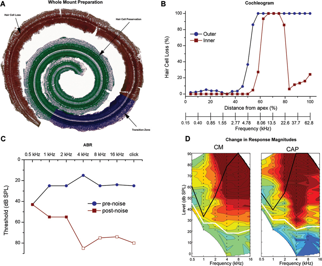
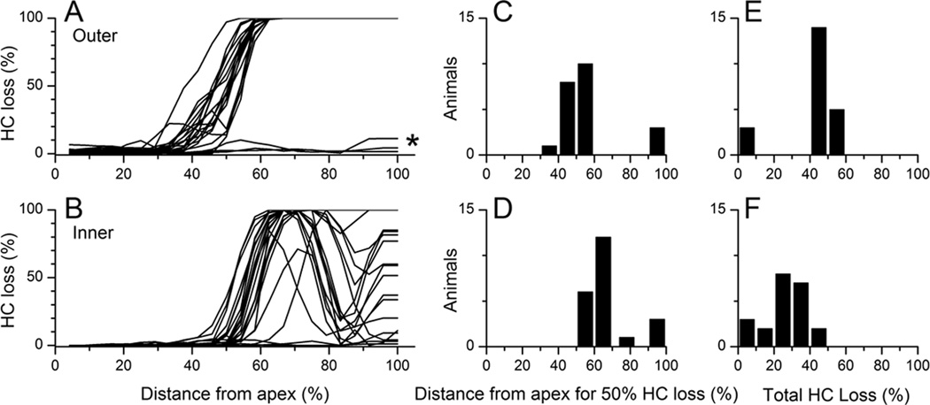
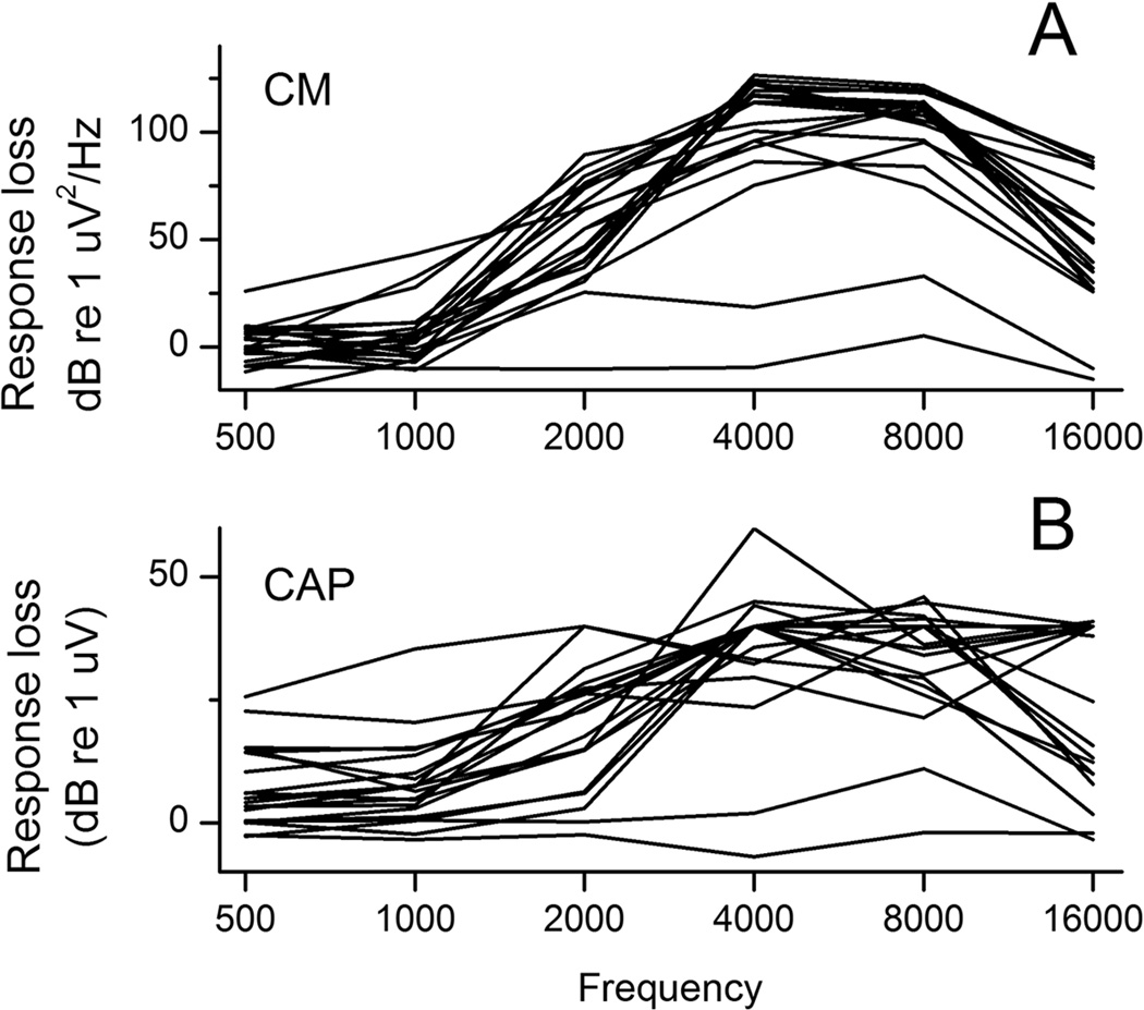
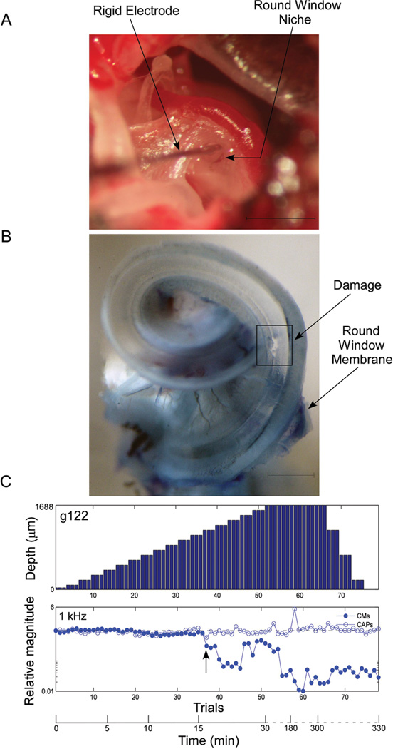
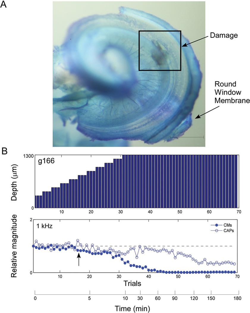
Methods: Gerbils underwent high-pass (4-kHz cutoff) noise exposure to produce sloping hearing loss. After 1 month of recovery, each animal's hearing loss was determined from auditory brainstem responses and baseline intracochlear recording of the cochlear microphonic and compound action potential (CAP) obtained at the round window. Subsequently, electrode insertions were performed to produce basal trauma, whereas the acoustically generated potentials to a 1-kHz tone-burst were recorded after each step of electrode advancement. Hair cell counts were made to characterize the noise damage, and cochlear whole mounts were used to identify cochlear trauma due to the electrode.

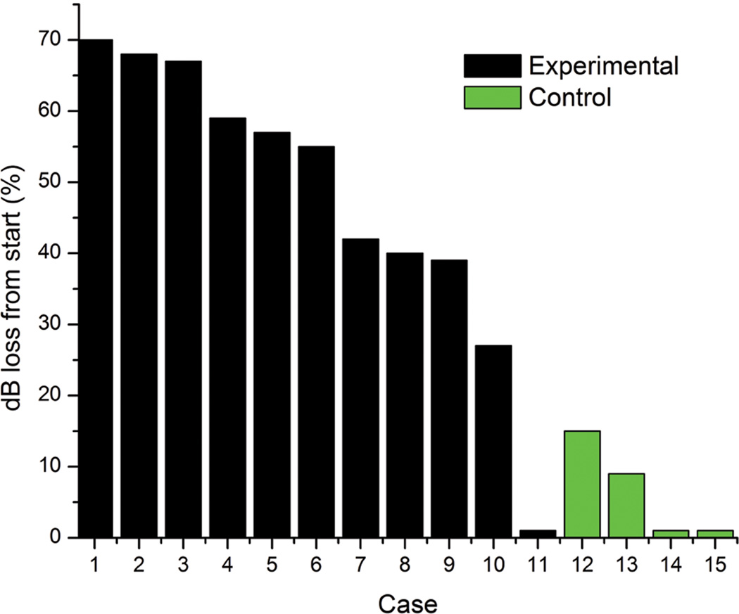
Results: The noise exposure paradigm produced a pattern of hair cell, auditory brainstem response, and intracochlear potential losses that closely mimicked that of electrical and acoustic stimulation patients. Trauma in the basal turn, in the 15- to 30-kHz portion of the deafened region, remote from preserved hair cells, induced a decline in intracochlear acoustic responses to the hearing preserved frequency of 1 kHz.

Conclusion: The results indicate that a recording algorithm based on physiological markers to low-frequency acoustic stimuli can identify cochlear trauma during implantation. Future work will focus on translating these results for use with current cochlear implant technology in humans.


Conflict of interest statement

Conflict of Interest: Dr. Oliver Adunka is a consultant for MED-EL Corporation and receives research support. Dr. Douglas Fitzpatrick receives research support from MED-EL Corporation. Dr. Craig Buchman serves as a consultant for Advanced Bionics, Cochlear Corp., and MED-EL Corporation.

Figures

Figure 1
Figure 1
A: Dissected BM-organ of Corti complex after noise exposure stained with cresyl violet. Three zones were identified under direct visualization and colored with photoshop. The cochlea shows a complete loss of OHCs in the basal region (red) followed by a transition zone with partial OHC loss (blue) and an apical area of complete OHC preservation (green). B: Cochleogram of inner and outer hair cell loss as a function of cochlear distance and frequency as determined from a gerbil cochlear frequency map (Mueller et al., 1996). C: Pre and Post noise exposure ABRs. Open symbols indicate no response at the highest intensity measured. D: Contour plots of the difference between the standard response at the round window of a normal hearing gerbil and the test response at the round window of a noise exposed gerbil. Green is no difference, blue is an increased response and red is a decreased response compared to normal hearing animals used as a standard (color scale of the CM is from 25 (dark blue) to 160 dB (red) re 1 µV/Hz, and the CAP is from 25 to 60 dB, re 1 µV). White lines are the thresholds of the standard and black lines are the thresholds of the test animal. There is a large response decrease for the higher frequencies (4 – 16 kHz) at the high intensities. There is small to no change in response seen to the high frequencies at the lower intensities since the standard response at these levels are small to none, therefore the magnitude of change is minimal to none even if there is no response in the noise exposed animals to these low levels.
Figure 2
Figure 2
A: OHC cochleograms for all noise exposed animals (n=22). Counts were made in 250 µm sections, and losses are in reference to average results in normal-hearing animals (n=3). Gerbil cochlear length was ~12 mm in all animals. B: IHC cochleograms. C and D: Histograms of the position of 50% hair cell loss as a function of distance along the BM. E and F. Histograms of total amount of hair cell loss.
Figure 3
Figure 3
Test of hearing loss due to noise exposure based on pre and post exposure ABRs to different frequency tone pips and click stimuli as indicated (n=20). Closed circles demonstrate measurable post-exposure thresholds while open circles indicate a minimum change based on the highest threshold tested.
Figure 4
Figure 4
The magnitude of response loss of the CM (A) and CAP (B) across frequencies relative to a normal hearing standard animal. These plots are essentially summing the columns of contour plots such as those shown in Fig. 1D.
Figure 5
Figure 5
Example of an intracochlear electrode penetration (case 122) with rapid decline in signals in the setting of sloping noise induced sensorineural hearing loss. A: Surgical situs demonstrating the exposed bulla including the round window. The electrode is a 50 µm tungsten rod as described. The electrode is directed towards the BM as evidenced by the darker stain of the stria vascularis observed through the round window membrane. Scale represents 1 mm. B: Cochlea whole mount specimen obtained after the animal was sacrificed. Round window membrane is preserved during dissection. Damage is observed in the basilar membrane. Scale represents 0.5 mm. C: Serial intracochlear recordings during electrode penetration. After each electrode advancement (between 50–100 µm), a response to one suprathreshold stimulus at 1,000 Hz was obtained and CM and CAP magnitudes were calculated. Then, these were compared to the standard obtained at the round window. Arrow indicates start of decline in CM response. A maximum insertion of 1688 um is noted, which is greater than most other insertions because we were confident the electrode had not penetrated into the second turn (later confirmed by histology). We took a more conservative approach in subsequent experiments with the shallower insertion depth of 1300 um.
Figure 6
Figure 6
Example of an intracochlear electrode penetration with gradual decline in response magnitudes in the setting of sloping noise induced sensorineural hearing loss. A: Cochlea whole mount specimen obtained after the animal was sacrificed. Note area of damage to the basilar membrane and also a large area of injury evidenced by presence of a blood clot on the osseous spiral lamina, medial to the basilar membrane damage. Scale represents 0.5 mm. B: Serial intracochlear recordings during electrode penetration as in Fig 5. Arrow indicates start of decline in CM response.
Figure 7
Figure 7
Histogram of percent CM response loss to 1 kHz tone burst for each electrode insertion case. Black bars represent 11 experimental cases where electrode was inserted to a depth of 1.3 – 1.7 mm past the round window. Green bars represent four control cases where electrode was inserted just past the round window to a depth of 0.4 mm.

References

    1. Gifford RH, Dorman MF, Shallop JK, Sydlowski SA. Evidence for the expansion of adult cochlear implant candidacy. Ear Hear. 2010;31:186–194. - PMC - PubMed
    1. Cullen RD, Higgins C, Buss E, Clark M, Pillsbury HC, 3rd, Buchman CA. Cochlear implantation in patients with substantial residual hearing. Laryngoscope. 2004;114:2218–2223. - PubMed
    1. Hodges AV, Schloffman J, Balkany T. Conservation of residual hearing with cochlear implantation. Am J Otol. 1997;18:179–183. - PubMed
    1. von Ilberg C, Kiefer J, Tillein J, et al. Electric-acoustic stimulation of the auditory system. New technology for severe hearing loss. ORL J Otorhinolaryngol Relat Spec. 1999;61:334–340. - PubMed
    1. Gantz BJ, Turner C. Combining acoustic and electrical speech processing: Iowa/Nucleus hybrid implant. Acta Otolaryngol. 2004;124:344–347. - PubMed

Publication types

MeSH terms