. 2011 Oct 28;12(11):1196-203.
doi: 10.1038/embor.2011.177.

Ixodes scapularis salivary gland protein P11 facilitates migration of Anaplasma phagocytophilum from the tick gut to salivary glands

Affiliations

Ixodes scapularis salivary gland protein P11 facilitates migration of Anaplasma phagocytophilum from the tick gut to salivary glands

Lei Liu et al. EMBO Rep. .

Abstract

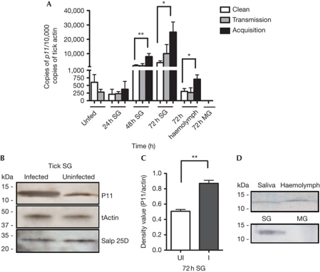
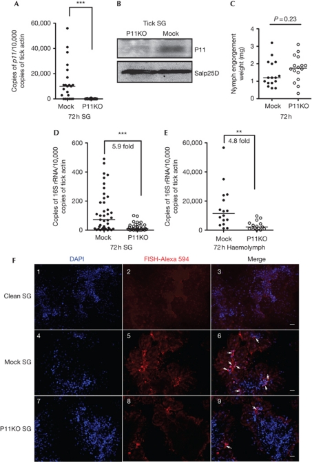
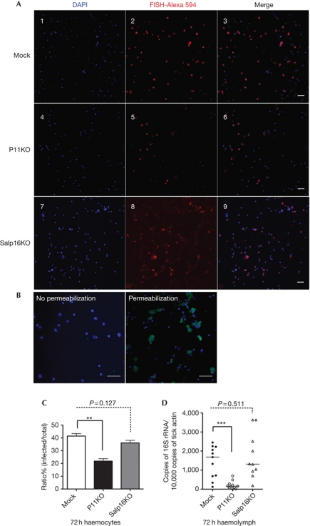
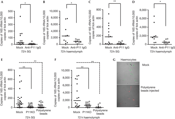
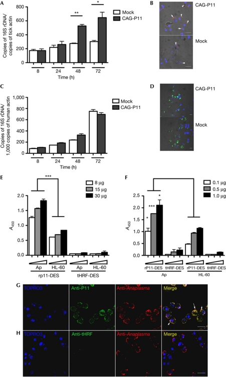
Ixodes ticks harbour several human pathogens belonging to the order Rickettsiales, including Anaplasma phagocytophilum, the agent of human anaplasmosis. When ticks feed on A. phagocytophilum-infected mice, the pathogen enters the ticks' gut. The bacteria then migrate from the gut to infect the salivary glands of the ticks and are transmitted to the next host via the saliva. The molecular mechanisms that enable the migration of A. phagocytophilum from the gut to the salivary glands are poorly understood. Here we show that a secreted tick protein, P11, is important in this process. We show that P11 enables A. phagocytophilum to infect tick haemocytes, which are required for the migration of A. phagocytophilum from the gut to the salivary glands. Silencing of p11 impaired the A. phagocytophilum infection of tick haemocytes in vivo and consequently decreased pathogen infection of the salivary glands. In vitro experiments showed that P11 could bind to A. phagocytophilum and thus facilitate its infection of tick cells. This report provides new insights into A. phagocytophilum infection of ticks and reveals new avenues to interrupt the life cycle of Anaplasma and related Rickettsial pathogens.


Conflict of interest statement

The authors declare that they have no conflict of interest.

Figures

Figure 1
Figure 1
Tick SG protein P11 is induced during tick feeding on A. phagocytophilum-infected mice. (A) The expression profile of p11 in unfed nymph ticks, or in the SG of naive nymphal ticks feeding on clean mice (Clean); A. phagocytophilum-infected mice (Acquisition); or A. phagocytophilum-infected nymphal ticks feeding on naive mice (Transmission); p11 expression levels in the haemolymph of nymphs during acquisition and transmission. (BD) Western blot assessment of P11 expression: during acquisition in tick SG (B,C); and in the haemolymph and MG of nymphal ticks during acquisition and in saliva from adult ticks (D). Tick actin and another unrelated tick SG protein Salp25D were used as loading control. Error bars show means±s.e.m. *P<0.05 and **P<0.01. The three independent experiments yielded similar results. MG, midgut; SG, salivary gland.
Figure 2
Figure 2
Silencing p11 expression decreases A. phagocytophilum burden in tick SG and haemolymph. (A) Silencing of p11 expression. (B) Specificity of p11 silencing at the protein level. Tick SG protein Salp25D was used as control. (C) Tick feeding was not impaired after silencing p11 expression. (D,E) The levels of A. phagocytophilum 16S rRNA transcripts in tick SG (D) and haemolymph (E). (F) RNA-FISH microscopy of A. phagocytophilum in clean tick SG (Clean SG, panels 1–3), in mock tick SG (Mock SG, panels 4–6) and in P11-deficient tick SG (P11KO SG, panels 7–9). The horizontal line represents the median. **P<0.01 and ***P<0.001. The three independent experiments yielded similar results. Scale bar, 50 μm. MG, midgut; RNA-FISH, RNA-fluorescence in situ hybridization; SG, salivary gland.
Figure 3
Figure 3
P11 is required for A. phagocytophilum infection of tick haemocytes. (A) (Panels 1–3) RNA-FISH images of A. phagocytophilum in mock group haemocytes (Mock); (panels 4–6) P11-deficient tick haemocytes (P11KO); (panels 7–9) Salp16-deficient tick haemocytes (Salp16KO). (B) A. phagocytopholum was detected in permeabilized tick haemocytes, but not in unpermeabilized haemocytes, by immunofluorescence analysis. (C) Quantification of A. phagocytophilum infection ratio in mock (Mock), P11-deficient (P11KO) and Salp16-deficient (Salp16KO) tick haemocytes. (D) Levels of A. phagocytophilum 16S rRNA transcripts in mock, p11-deficient (p11KO) and salp16-deficient (Salp16KO) tick haemolymph. Error bars show means±s.e.m. The horizontal line represents the median. **P<0.01 and ***P<0.001. The three independent experiments yielded similar results. Scale bar, 50 μm. RNA-FISH, RNA-fluorescence in situ hybridization.
Figure 4
Figure 4
A. phagocytophilum migration in tick SG and haemolymph was blocked by P11 antiserum and polystyrene beads. (A,B) Normal rabbit IgG (Mock) or purified rabbit anti-P11 IgG was injected into the haemocoel and ticks fed on A. phagocytophilum-infected mice. The levels of A. phagocytophilum 16S rRNA transcripts were compared in SG (A) and haemolymph (B). (C,D) A. phagocytophilum-infected mice were passively immunized by normal rabbit IgG (Mock) or purified rabbit anti-P11 IgG (P11 Ab), and the levels of A. phagocytophilum 16S rRNA were measured in tick SG (C) and haemolymph (D) after feeding for 72 h. (E,F) Polystyrene beads were injected into the haemocoel of clean nymphs and the ticks fed on A. phagocytophilum-infected mice. The levels of A. phagocytophilum 16S rRNA transcripts were compared in SG (E) and haemolymph (F). (G) Confocal microscopy image of A. phagocytophilum (Alexa488) in mock and polystyrene beads-injected haemocytes (red amine labelled). The horizontal line represents the median. *P<0.05 and **P<0.01. The three independent experiments yielded similar results. Scale bar, 50 μm. SG, salivary gland.
Figure 5
Figure 5
P11 facilitate A. phagocytophilum infection by binding to the bacteria. (A,C) The levels of A. phagocytophilum 16S rRNA transcripts at different time points after infection of control (Mock) and p11-overexpressed (CAG-P11) I. ricinus tick cells (A) and HL-60 cells (C). (B,D) Confocal microscopy of A. phagocytophilum in control (Mock) and p11-overexpressed (CAG-P11) (B) I. ricinus tick cells and (D) HL-60 cells. (E,F) Dose-dependent increase in binding of the A. phagocytophilum-enriched fraction to rP11-DES (E), and that of rP11-DES to the A. phagocytophilum-enriched fraction (F). Tick salivary protein, tHRF, was used as control. (G,H) Confocal microscopy of infected tick cells shows direct interaction between P11 and A. phagocytophilum (G); tHRF was used as control (H). Error bars show means±s.e.m. *P<0.05, **P<0.01 and ***P<0.001. The three independent experiments yielded similar results. Scale bar, 20 μm.

Similar articles

Cited by

References

    1. Azad AF, Beard CB (1998) Rickettsial pathogens and their arthropod vectors. Emerg Infect Dis 4: 179–186 - PMC - PubMed
    1. Bakken JS, Dumler JS (2000) Human granulocytic ehrlichiosis. Clin Infect Dis 31: 554–560 - PubMed
    1. Bakken JS, Dumler JS, Chen SM, Eckman MR, Van Etta LL, Walker DH (1994) Human granulocytic ehrlichiosis in the upper Midwest United States. A new species emerging? JAMA 272: 212–218 - PubMed
    1. Borovickova B, Hypsa V (2005) Ontogeny of tick hemocytes: a comparative analysis of Ixodes ricinus and Ornithodoros moubata. Exp Appl Acarol 35: 317–333 - PubMed
    1. Carlyon JA, Fikrig E (2006) Mechanisms of evasion of neutrophil killing by Anaplasma phagocytophilum. Curr Opin Hematol 13: 28–33 - PubMed

Publication types

MeSH terms

Substances