. 2011 Dec;18(12):884-96.
doi: 10.1038/cgt.2011.57. Epub 2011 Sep 16.

Antitumor effect of sFlt-1 gene therapy system mediated by Bifidobacterium Infantis on Lewis lung cancer in mice

Affiliations
Free PMC article

Antitumor effect of sFlt-1 gene therapy system mediated by Bifidobacterium Infantis on Lewis lung cancer in mice

H Zhu et al. Cancer Gene Ther. 2011 Dec.
Free PMC article

Abstract

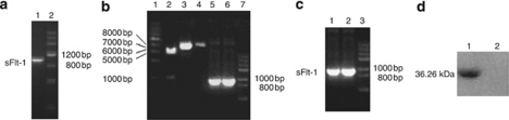
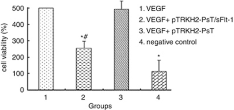
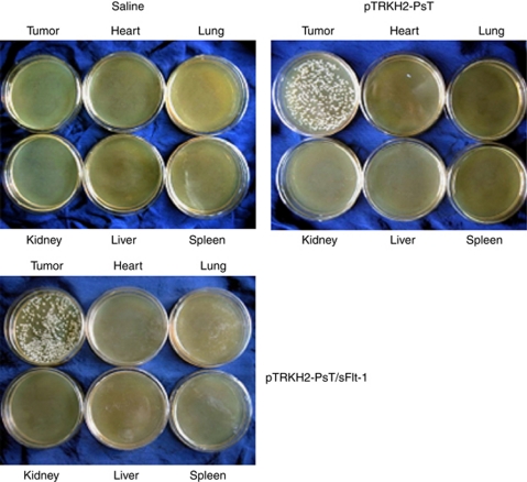
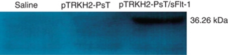
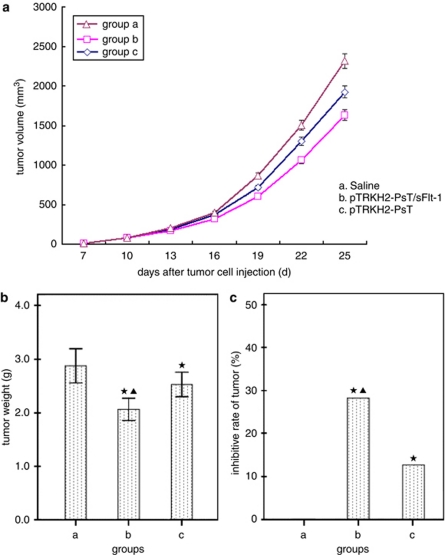
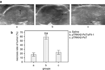
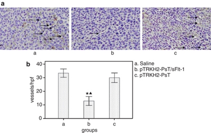
Soluble fms-like tyrosine kinase receptor (sFlt-1) is a soluble form of extramembrane part of vascular endothelial growth factor receptor-1 (VEGFR-1) that has antitumor effects. Bifidobacterium Infantis is a kind of non-pathogenic and anaerobic bacteria that may have specific targeting property of hypoxic environment inside of solid tumors. The aim of this study was to construct Bifidobacterium Infantis-mediated sFlt-1 gene transferring system and investigate its antitumor effect on Lewis lung cancer (LLC) in mice. Our results demonstrated that the Bifidobacterium Infantis-mediated sFlt-1 gene transferring system was constructed successfully and the system could express sFlt-1 at the levels of gene and protein. This system could not only significantly inhibit growth of human umbilical vein endothelial cells induced by VEGF in vitro, but also inhibit the tumor growth and prolong survival time of LLC C57BL/6 mice safely. These data suggest that Bifidobacterium Infantis-mediated sFlt-1 gene transferring system presents a promising therapeutic approach for the treatment of cancer.


Figures

Figure 1
Figure 1
The results of electrophoresis and western blot. (a) Polymerase chain reaction (PCR) products of soluble fms-like tyrosine kinase receptor (sFlt-1). Lane 1: sFlt-1; lane 2: DNA marker III. (b) Digestion of sFlt-1 and pTRKH2-PsT by restriction endonuclease and PCR products of sFlt-1. Lane 1: 1 kb DNA ladder; lane 2: recombinant plasmid pTRKH2-PsT/sFlt-1 digested by restriction endonuclease BamHI and SalI; lane 3: recombinant plasmid pTRKH2-PsT/sFlt-1; lane 4: plasmid pTRKH2-PsT; lane 5: PCR products of sFlt-1 gene from recombinant plasmid pTRKH2-PsT/sFlt-1; lane 6: PCR products of sFlt-1 gene from recombinant plasmid pcDNA3.1/sFlt-1; lane 7: DNA marker III (TransGen). (c) Agarose gel electrophoresis of reverse transcription (RT)-PCR and PCR products from recombinant Bifidobacterium Infantis of pTRKH2-PsT/sFlt-1. Lane 1: PCR products of sFlt-1 gene from recombinant plasmid pTRKH2-PsT/sFlt-1; lane 2: RT-PCR product of RNA purified from Bifidobacterium Infantis with pTRKH2-PsT/sFlt-1; lane 3: DNA marker III (from TransGen). (d) Western blot analysis of the expression of sFlt-1 gene. A western blot band (36.26 kDa) from Bifidobacterium Infantis transformed recombinant pTRKH2-PsT/sFlt-1 plasmid was shown (lane 1), and no same band was shown from Bifidobacterium Infantis transformed recombinant pTRKH2-PsT (lane 2).
Figure 2
Figure 2
Effect of Bifidobacterium Infantis-mediated pTRKH2-PsT/sFlt-1 delivery system on HUVECs. 1: VEGF; 2: VEGF and treated fluid of the Bifidobacterium Infantis transformed with recombinant pTRKH2-PsT/sFlt-1 plasmid; 3: VEGF and treated fluid of the Bifidobacterium Infantis transformed with pTRKH2-PsT plasmid; 4: negative control. *P<0.05 vs VEGF group; #P<0.05 vs negative control.
Figure 3
Figure 3
Analysis of targeting property of Bifidobacterium Infantis. Saline: saline control group; pTRKH2-PsT: recombinant Bifidobacterium Infantis containing pTRKH2-PsT plasmid group; pTRKH2-PsT/sFlt-1: recombinant Bifidobacterium Infantis containing pTRKH2-PsT/sFlt-1 plasmid group.
Figure 4
Figure 4
Western blot analysis of the expression of sFlt-1 gene in tumor tissue. Saline: saline control group; pTRKH2-PsT: recombinant Bifidobacterium Infantis containing pTRKH2-PsT plasmid group; pTRKH2-PsT/sFlt-1: recombinant Bifidobacterium Infantis containing pTRKH2-PsT/sFlt-1 plasmid group. The western blot showed that a band of about 36.26 kDa from Bifidobacterium Infantis transformed recombinant pTRKH2-PsT/sFlt-1 plasmid was shown, and no same band was shown from Bifidobacterium infantis transformed recombinant pTRKH2-PsT or saline control group.
Figure 5
Figure 5
Effect of Bifidobacterium Infantis-mediated pTRKH2-PsT/sFlt-1 delivery system on the growth of Lewis lung cancer C57BL/6 mice. (a) Tumor growth curve; (b) tumor weight; and (c) inhibitive rate of tumor. a: saline control group and b: recombinant Bifidobacterium Infantis containing pTRKH2-PsT/sFlt-1 plasmid group; c: recombinant Bifidobacterium Infantis containing pTRKH2-PsT plasmid group. *P<0.05 vs group a; P<0.05 vs group c.
Figure 6
Figure 6
Effect of Bifidobacterium Infantis-mediated pTRKH2-PsT/sFlt-1 delivery system on tumor necrosis. (a) Ultrasound images of tumor and (b) necrosis rate of tumor. a: saline control group; b: recombinant Bifidobacterium Infantis containing pTRKH2-PsT/sFlt-1 plasmid group; and c: recombinant Bifidobacterium Infantis containing pTRKH2-PsT plasmid group. Arrows showed the necrotic area. *P<0.05 vs group a; P<0.05 vs group c.
Figure 7
Figure 7
Observation of blood flow in tumors by color Doppler flow imaging (CDFI). (a) Blood flow image of tumor and (b) classification of blood flow signals. a: saline control group; b: recombinant Bifidobacterium Infantis containing pTRKH2-PsT/sFlt-1 plasmid group; and c: recombinant Bifidobacterium Infantis containing pTRKH2-PsT plasmid group. Grade: 0, no blood flow signals detected within the tumor; I, minimal blood flow (one or two dot-like or a thin- and short-like blood flow signals detected within the tumor); II, moderate blood flow (up to three dot-like blood flow signals or one longer blood flow signals detected within the tumor); III, abundant blood flow (more than five dot-like blood flow signals or two longer blood flow signals detected within the tumor).
Figure 8
Figure 8
Detection of the tumor microvessel density (MVD) by CD31 immunohistochemistry. (a) Photomicrograph of immunohistochemical staining of CD31 ( × 200) and (b) MVD. a: Saline control group; b: recombinant Bifidobacterium Infantis containing pTRKH2-PsT/sFlt-1 plasmid group; and c: recombinant Bifidobacterium Infantis containing pTRKH2-PsT plasmid group. *P<0.05 vs group a; P<0.05 vs group c. Arrows direct to the vessels.
Figure 9
Figure 9
Survival curves of Lewis lung cancer C57BL/6 mice. a: Saline control group; b: recombinant Bifidobacterium Infantis containing pTRKH2-PsT/sFlt-1 plasmid group; and c: recombinant Bifidobacterium Infantis containing pTRKH2-PsT plasmid group.

Similar articles

Cited by

References

    1. Sonmezer M, Gungor M, Ensari A, Ortac F. Prognostic significance of tumor angiogenesis in epithelial ovarian cancer: in association with transforming growth factor [beta] and vascular endothelial growth factor. Int J Gynecol Cancer. 2004;14:82–88. - PubMed
    1. Li W, Xu RJ, Zhang HH, Jiang LH. Overexpression of cyclooxygenase-2 correlates with tumor angiogenesis in endometrial carcinoma. Int J Gynecol Cancer. 2006;16:1673–1678. - PubMed
    1. Cantu De Leon D, Lopez-Graniel C, Mendivil MF, Vilchis GC, Gomez C, Salazar JDLG. Significance of microvascular density (MVD) in cervical cancer recurrence. Int J Gynecol Cancer. 2003;13:856–862. - PubMed
    1. Chen WT, Huang CJ, Wu MT, Yang SF, Su YC, Chai CY. Hypoxia-inducible factor-1[alpha] is associated with risk of aggressive behavior and tumor angiogenesis in gastrointestinal stromal tumor. Jpn J Clin Oncol. 2005;35:207–213. - PubMed
    1. Joo YEMD, Rew JSMD, Seo YHMD, Choi SKMD, Kim YJMD, Park CSMD, et al. Cyclooxygenase-2 overexpression correlates with vascular endothelial growth factor expression and tumor angiogenesis in gastric cancer. J Clin Gastroenterol. 2003;37:28–33. - PubMed

Publication types

Substances