. 2011:17:2283-91.
Epub 2011 Aug 25.

Structure of corneal layers, collagen fibrils, and proteoglycans of tree shrew cornea

Affiliations

Structure of corneal layers, collagen fibrils, and proteoglycans of tree shrew cornea

Turki Almubrad et al. Mol Vis. 2011.

Abstract

Purpose: The stroma is the major part of the cornea, in which collagen fibrils and proteoglycans are distributed uniformly. We describe the ultrastructure of corneal layers, collagen fibrils (CF), and proteoglycans (PGs) in the tree shrew cornea.

Methods: Tree shrew corneas (5, 6, and 10 week old animals) and normal human corneas (24, 25, and 54 years old) were fixed in 2.5% glutaraldehyde containing cuprolinic blue in a sodium acetate buffer. The tissue was processed for electron microscopy. The 'iTEM Olympus Soft Imaging Solutions GmbH' program was used to measure the corneal layers, collagen fibril diameters and proteoglycan areas.

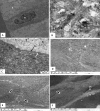
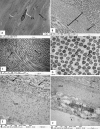
Results: The tree shrew cornea consists of 5 layers: the epithelium, Bowman's layer, stroma, Descemet's membrane, and endothelium. The epithelium was composed of squamous cells, wing cells and basal cells. The Bowman's layer was 5.5±1.0 µm thick and very similar to a normal human Bowman's layer. The stroma was 258±7.00 µm thick and consisted of collagen fibril lamellae. The lamellae were interlaced with one another in the anterior stroma, but ran parallel to one another in the middle and posterior stroma. Collagen fibrils were decorated with proteoglycan filaments with an area size of 390 ±438 nm(2). The collagen fibril had a minimum diameter of 39±4.25 nm. The interfibrillar spacing was 52.91±6.07 nm. Within the collagen fibrils, very small electron-dense particles were present.

Conclusions: The structure of the tree shrew cornea is very similar to that of the normal human cornea. As is the case with the human cornea, the tree shrew cornea had a Bowman's layer, lamellar interlacing in the anterior stroma and electron-dense particles within the collagen fibrils. The similarities of the tree shrew cornea with the human cornea suggest that it could be a good structural model to use when studying changes in collagen fibrils and proteoglycans in non-genetic corneal diseases, such as ectasia caused after LASIK (laser-assisted in situ keratomileusis).


Figures

Figure 1
Figure 1
Light micrograph of tree shrew cornea. A: Tree shrew cornea consisting of epithelium, Bowman’s layer, stroma, Descemet’s membrane, and endothelium. Note the structure of the cornea is very similar to the human cornea and shows 5 layers. B: Part of the tree shrew cornea showing squamous cells, wing cells, basal epithelial cells and Bowman’s layer. C: Part of the corneal stroma containing keratocytes. D: Part of the cornea showing Descemet’s membrane and endothelium. B=Bowman’s layer, E=Epithelium, DM=Descemet’s membrane, KR=Keratocyte, EN=endothelium, and S=Stroma.
Figure 2
Figure 2
Electron micrograph of tree shrew and human cornea fixed in 2.5% glutaraldehyde containing cuprolinic blue and embedded in spurr resin. A: Basal epithelial cells are columnar and contain large nuclei. B: Prominent cytoplasmic filaments in basal epithelial cells. C: Basal epithelial cells attached by hemidesmosomes to a basement membrane followed by a Bowman’s layer consisting of dense, randomly arranged collagen fibrils. D: Non linear, random distribution of collagen fibrils (curved arrowhead) present in the anterior stroma just below the Bowman's layer. Some of the collagen run across the longitudinally-running collagen fibrils (arrowhead). E: Lamellae are interlacing (arrowhead) in the anterior stroma of the tree shrew. F: Lamellae are interlacing (arrowhead) in the anterior stroma of the normal human cornea. B=Bowman’s layer, BM=Basement membrane, CF=Collagen fibrils, E=Epithelium, H=Hemidesmosomes, and MF=Cytoplasmic filaments.
Figure 3
Figure 3
Electron micrograph of tree shrew cornea fixed in 2.5% glutaraldehyde containing cuprolinic blue and embedded in spurr resin. A: In the middle stroma, parallel running lamellae containing a keratocyte. B and C: Lamella containing orderly, packed collagen fibrils and proteoglycans. D: In cross-section, collagen fibrils exhibiting tiny particles, some of which are of high electron density. E: Pre-Descemet's stroma containing very fine fibrils and large PGs around the collagen fibrils. Fibrillar structures are present throughout the Descemet's membrane. F: Part of the posterior cornea, showing a banded Descemet's membrane and an endothelium containing a nucleus; also a prominent endoplasmic reticulum. CF=Collagen fibrils, DM=Descemet's membrane, EN=endothelium, F=Fine fibril, G=tiny particles, KR=Keratocytes, L=Lamella, PG=Proteoglycan, and S=Stroma.
Figure 4
Figure 4
Electron micrograph of tree shrew and human cornea fixed in 2.5% glutaraldehhyde containing cuprolinic blue and embedded in spurr resin. A: Electron micrograph of collagen fibrils of tree shrew cornea. B: Digital image obtained after processing the image shown in A. C: Electron micrograph of collagen fibrils of human cornea. D: Digital image obtained after processing the image shown in C. The images were displayed by using color coding to demonstrate the distribution of the variable diameters of collagen fibrils. Collagen fibril color coding: Red=15–20 nm, Green=20–25 nm, Blue=25–30 nm, Yellow=30–35 nm, Aqua=35–40 nm, Pink=40–45 nm, and Brown=45–50 nm.
Figure 5
Figure 5
Electron micrograph of tree shrew and human cornea fixed in 2.5% glutaraldehhyde containing cuprolinic blue and embedded in spurr resin. A: Electron micrograph of proteoglycans of tree shrew stroma. B: Digital image obtained after processing image shown in A, showing variable area distribution of PGs. C: Electron micrograph of proteoglycans of human stroma. D: Digital image obtained after processing image shown in C, showing variable area distribution of PGs. PG=Proteoglycan; Proteoglycans color coding: Red=50–350 nm2, Green=350–650 nm2, Blue=650–950 nm2, Yellow=950–1,250 nm2, Aqua=1,250–1,550 nm2, Pink=1,550–1,850 nm2, and Brown=1,850–2,150 nm2.

Similar articles

Cited by

References

    1. Siegwart JT, Jr, Norton TT. The susceptible period for deprivation-induced myopia in tree shrews. Vision Res. 1998;38:3505–15. - PubMed
    1. Norton TT, Amedo AO, John T. Siegwart Jr. Darkness causes myopia in visually experienced tree shrews. Invest Ophthalmol Vis Sci. 2006;47:4700–7. - PMC - PubMed
    1. McBrien NA, Norton TT. Prevention of collagen crosslinking increases form-deprivation myopia in tree shrews. Exp Eye Res. 1994;59:475–86. - PubMed
    1. Jobling AI, Nguyen M, Gentle A, McBrien NA. Isoform-specific changes in scleral transforming growth factor-beta expression and the regulation of collagen synthesis during myopia progression. J Biol Chem. 2004;279:18121–6. - PubMed
    1. Ishiko S, Yoshida A, Mori F, Abiko T, Kitaya N, Kojima M, Saito K. Early ocular changes in a tree shrew model of diabetes. Nippon Ganka Gakkai Zasshi. 1997;101:19–23. - PubMed

Publication types

LinkOut - more resources