

Structure of corneal layers, collagen fibrils, and proteoglycans of tree shrew cornea
- PMID: 21921979
- PMCID: PMC3171502
Structure of corneal layers, collagen fibrils, and proteoglycans of tree shrew cornea
Abstract
Purpose: The stroma is the major part of the cornea, in which collagen fibrils and proteoglycans are distributed uniformly. We describe the ultrastructure of corneal layers, collagen fibrils (CF), and proteoglycans (PGs) in the tree shrew cornea.
Methods: Tree shrew corneas (5, 6, and 10 week old animals) and normal human corneas (24, 25, and 54 years old) were fixed in 2.5% glutaraldehyde containing cuprolinic blue in a sodium acetate buffer. The tissue was processed for electron microscopy. The 'iTEM Olympus Soft Imaging Solutions GmbH' program was used to measure the corneal layers, collagen fibril diameters and proteoglycan areas.
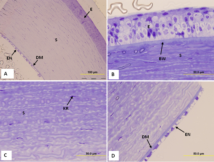
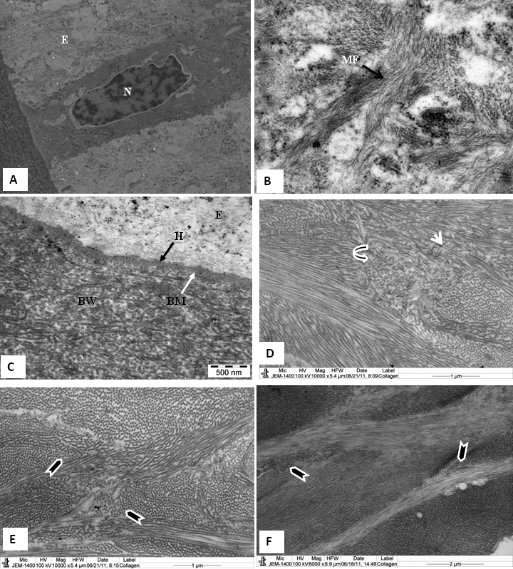
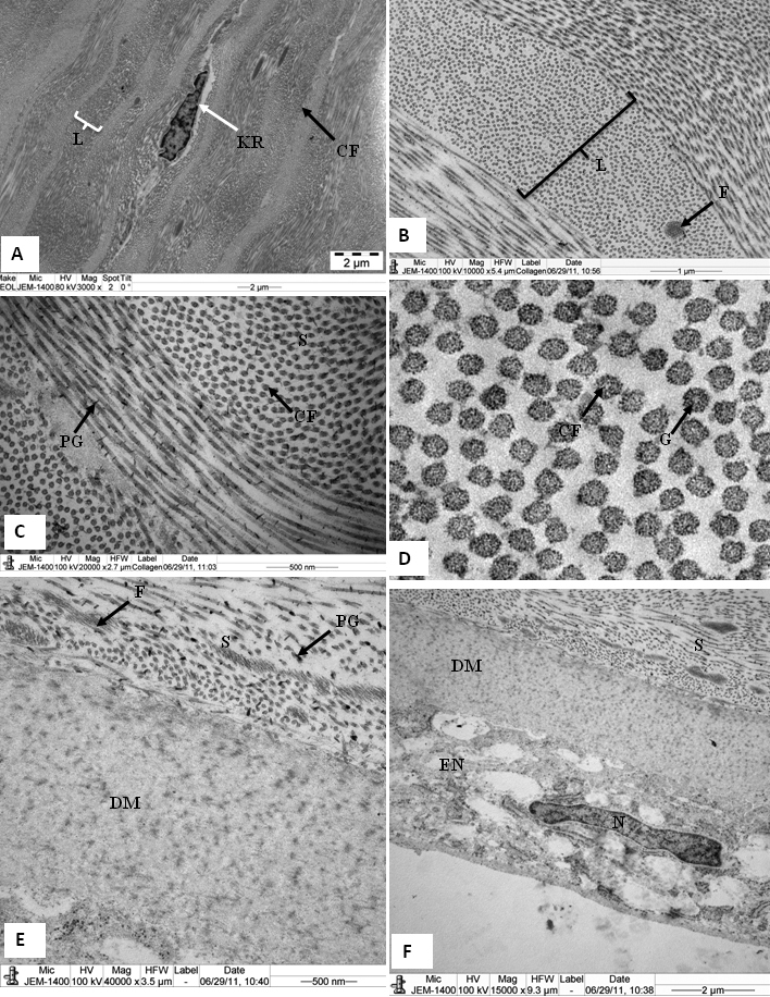
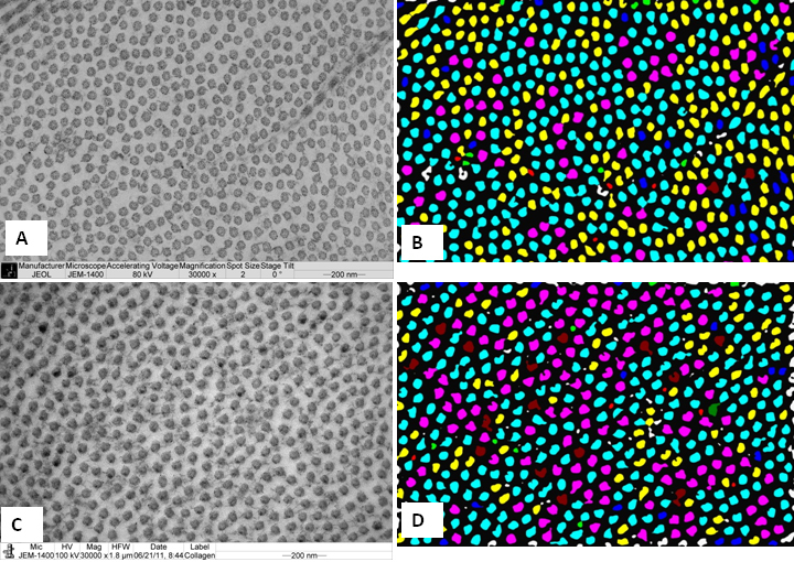
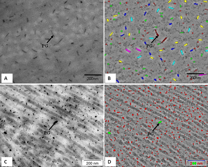
Results: The tree shrew cornea consists of 5 layers: the epithelium, Bowman's layer, stroma, Descemet's membrane, and endothelium. The epithelium was composed of squamous cells, wing cells and basal cells. The Bowman's layer was 5.5±1.0 µm thick and very similar to a normal human Bowman's layer. The stroma was 258±7.00 µm thick and consisted of collagen fibril lamellae. The lamellae were interlaced with one another in the anterior stroma, but ran parallel to one another in the middle and posterior stroma. Collagen fibrils were decorated with proteoglycan filaments with an area size of 390 ±438 nm(2). The collagen fibril had a minimum diameter of 39±4.25 nm. The interfibrillar spacing was 52.91±6.07 nm. Within the collagen fibrils, very small electron-dense particles were present.
Conclusions: The structure of the tree shrew cornea is very similar to that of the normal human cornea. As is the case with the human cornea, the tree shrew cornea had a Bowman's layer, lamellar interlacing in the anterior stroma and electron-dense particles within the collagen fibrils. The similarities of the tree shrew cornea with the human cornea suggest that it could be a good structural model to use when studying changes in collagen fibrils and proteoglycans in non-genetic corneal diseases, such as ectasia caused after LASIK (laser-assisted in situ keratomileusis).
Figures
References
-
- Siegwart JT, Jr, Norton TT. The susceptible period for deprivation-induced myopia in tree shrews. Vision Res. 1998;38:3505–15. - PubMed
-
- McBrien NA, Norton TT. Prevention of collagen crosslinking increases form-deprivation myopia in tree shrews. Exp Eye Res. 1994;59:475–86. - PubMed
-
- Jobling AI, Nguyen M, Gentle A, McBrien NA. Isoform-specific changes in scleral transforming growth factor-beta expression and the regulation of collagen synthesis during myopia progression. J Biol Chem. 2004;279:18121–6. - PubMed
-
- Ishiko S, Yoshida A, Mori F, Abiko T, Kitaya N, Kojima M, Saito K. Early ocular changes in a tree shrew model of diabetes. Nippon Ganka Gakkai Zasshi. 1997;101:19–23. - PubMed
Publication types
MeSH terms
Substances
LinkOut - more resources
Full Text Sources
Research Materials
Miscellaneous