. 2011:17:2333-42.
Epub 2011 Aug 30.

Genotype-phenotype correlation of TGFBI corneal dystrophies in Polish patients

Affiliations

Genotype-phenotype correlation of TGFBI corneal dystrophies in Polish patients

Anna K Nowińska et al. Mol Vis. 2011.

Abstract

Purpose: To analyze genotype-phenotype correlation in patients originating from Polish population with the transforming growth factor beta induced (TGFBI) corneal dystrophies.

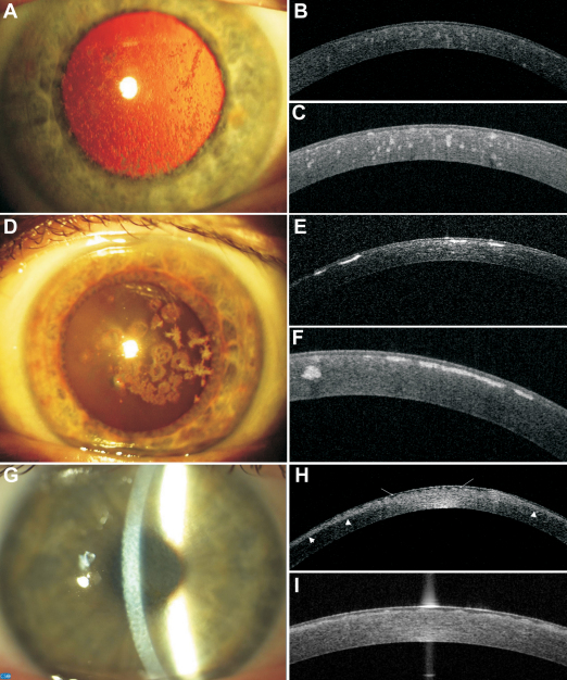
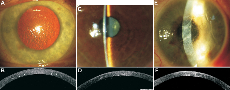
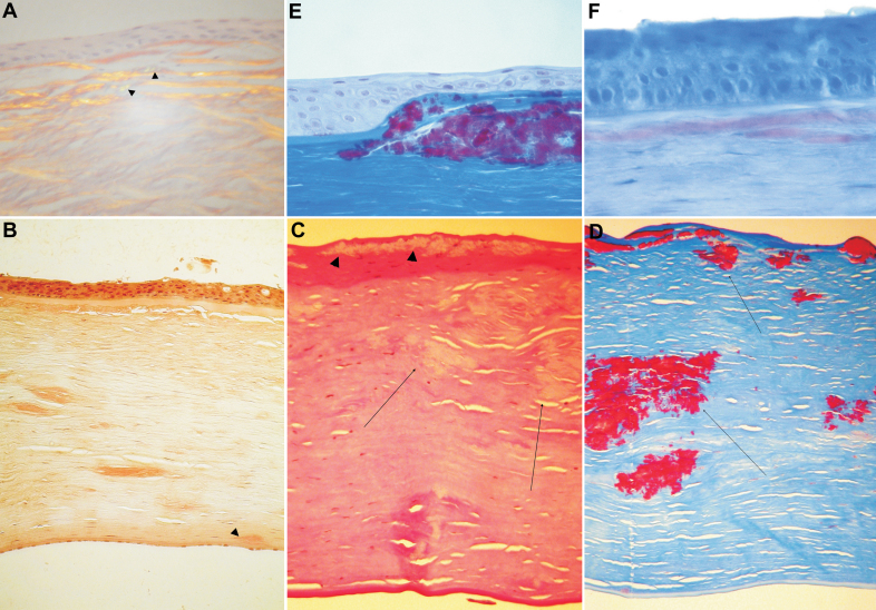
Methods: Sixty affected and 31 unaffected individuals from 15 unrelated Polish families were included in the study. The clinical diagnosis was based on the slit-lamp exam, 1310 nm time domain and 1310 nm swept source spectral domain optical coherence tomography (OCT). Histopathologic analysis was performed on 10 available corneal buttons. Exons of the TGFBI gene were screened for mutations with polymerase chain reaction (PCR) and direct DNA sequencing.

Results: We found the lattice phenotype dominant compared to the granular one in the Polish population (41:16 patients; lattice:granular). We identified five distinct mutations responsible for TGFBI corneal dystrophies (R124R, R124H, R555W, R555Q, and H626R). There was a strong genotype-phenotype correlation in the case of R124R and R555W mutations, while there was a distinct phenotypic heterogeneity in the case of the H626R mutation. OCT analysis revealed that the reflectivity, location and pattern of the corneal deposits were different among the TGFBI corneal dystrophies. The advantage of spectral swept source OCT over time-domain OCT scans is a more distinct visualization of the Bowman's layer area and deposits located under the epithelium.

Conclusions: This study underlines the role of comprehensive phenotype-genotype analysis in TGFBI corneal dystrophies, describes for the first time the TGFBI mutation spectrum in a Polish population and reveals phenotypic heterogeneity in the case of the H626R mutation.


Figures

Figure 1
Figure 1
Representative images of slit-lamp photographs, 1310 nm time-domain and 1310 nm swept source spectral domain optical coherence tomography scans of patients with granular corneal dystrophy type I (family F4), granular corneal dystrophy type II (family F9), and Thiel-Behnke corneal dystrophy (family F8). A: Female patient (F4; 53 years). Slit-lamp photograph showing gray-white granular deposits located centrally, with clear intervening stroma. GCDI/ R555W mutation. B: Female patient (F4; 53 years). High-resolution corneal scan – 1310 nm time. domain OCT. Focal granular hyperreflective changes located at different depths within the corneal stroma. GCDI/ R555W mutation. C: Female patient (F4; 53 years). Radial scan-swept source 1310 nm spectral OCT. Focal granular hyperreflective changes located at different depths within the corneal stroma. The Bowman’s layer area shows a distinct irregularity. GCDI/ R555W mutation. D: Female patient (F9; 44 years). Slit-lamp photograph. Centrally located, multiform: star- and disc-shaped opacities. No lattice lines are visible, either on direct light nor on retroillumination. GCDII/ R124H mutation. E: Female patient (F9; 44 years). High-resolution corneal scan – 1310 nm time domain OCT. Highly reflective opacities with distinct borders located in the anterior corneal part. GCDII/ R124H mutation. F: Female patient (F9; 44 years). Radial scan-swept source 1310 nm spectral OCT. Highly reflective disc-shaped changes located in the anterior stroma, under the epithelium, involving Bowman’s layer. One hyperreflective granular opacity located deeper in the mid stroma. GCDII/ R124H mutation. G: Female patient (F8; 38 years). Slit-lamp photograph. Diffuse corneal changes showing reticular, “honeycomb” pattern located in the anterior corneal part. TBCD/ R555Q mutation. H: Female patient (F8; 38 years). High-resolution corneal scan – 1310 nm time domain OCT. The diffuse boarder of increased reflectivity in the anterior part of the cornea (arrowheads). In the Bowman’s layer area, there is a distinct irregularity due to corneal opacities (arrows). TBCD/ R555Q mutation. I: Female patient (F8; 38 years). Radial scan-swept source 1310 nm spectral OCT. Bowman’s layer is replaced by an irregular pattern of opacities. TBCD/R555Q mutation.
Figure 2
Figure 2
Representative images of slit-lamp photographs and 1310 nm time-domain optical coherence tomography scans of patients with lattice corneal dystrophy type I (family F1); lattice corneal dystrophy variants (families F2 and F7). There is a noticeable phenotypic heterogeneity between corneal morphology of lattice corneal dystrophy variants caring the same H626R mutation. A: Male patient (F1; 37 years). Slit-lamp retroillumination photograph showing diffuse multiple lattice lines. LCDI/R124C mutation. B: Male patient (F1; 37 years). High-resolution corneal scan – 1310 nm time. domain OCT. There is a diffuse border between the anterior part of increased reflectivity and normal corneal stroma (arrowheads). The areas of increased stromal reflectivity correspond with corneal opacities. LCDI/R124C mutation. C: Female patient (F2; 45 years). Slit-lamp photograph. Delicate, fragile, rare lattice lines located centrally. LCD variant/ H626 mutation. D: Female patient (F2; 45 years). High-resolution corneal scan – 1310 nm time. domain OCT. Opacities with increased reflectivity visible through the whole depth of the cornea. Some of the opacities are located in the posterior corneal part (arrows). LCD variant/H626 mutation. E: Female patient (F7; 48 years). Slit-lamp photograph. Thick, distinct lines accompanied by stromal haze extended from limbus to limbus. LCD variant/H626 mutation. Note the distinct heterogeneity compared to Figure 2C. F: Female patient (F7; 48 years). High-resolution corneal scan – 1310 nm time. domain OCT. Opacities with increased reflectivity located mainly in the posterior corneal part causing distortion of the posterior corneal surface. LCD variant/H626 mutation.
Figure 3
Figure 3
Representative images of histopathologic analysis of four corneal sections – three after penetrating keratoplasty and one after deep anterior lamellar keratoplasty. A: Section of the cornea after deep anterior lamellar keratoplasty. Male patient (F1; 37 years old). Green birefringence is visible with a polarizing filter (arrowheads). Stromal deposition of amyloid substance in anterior corneal part distorts the architecture of the corneal lamellae. The absence of Bowman’s layer and thinning of the epithelium are noticeable. LCDI/R124C mutation. B: Section of the cornea after penetrating keratoplasty. Congo red stain. Female patient (F2; 45 years). Deposits throughout the corneal stroma stain positive with Congo red. Note the deep, posterior corneal location of the deposit (arrowhead). LCD variant/H626 mutation. C: Section of the cornea after penetrating keratoplasty. PAS stain. Female patient (F4; 53 years). Note the absence of Bowman’s layer and the distorted epithelium in correspondence of the granular deposits (arrowheads). There are several granular deposits throughout the corneal stroma (arrows). GCDI/R555W mutation. D: Section of the cornea after penetrating keratoplasty. PAS stain. Female patient (F4; 53 years). Masson trichrome stain. Section of the cornea showing the absence of Bowman’s layer and the absence of the epithelium in correspondence with the Masson trichrome – positive granular deposits (arrows). GCDI/R555W mutation. E: Section of the cornea after penetrating keratoplasty. Masson trichrome stain. Female patient (F9; 44 years). Note that the granular deposits are placed under the thinner epithelium, thus taking the place of the former Bowman’s layer. GCDII/R124H mutation. F: Section of the cornea after penetrating keratoplasty. Congo red stain. Female patient (F9; 44 years). Note the Congo red positive deposits in the anterior corneal stroma. GCDII/R124H mutation.

Similar articles

Cited by

References

    1. Weiss JS, Møller HU, Lisch W, Kinoshita S, Aldave AJ, Belin MW, Kivelä T, Busin M, Munier FL, Seitz B, Sutphin J, Bredrup C, Mannis MJ, Rapuano CJ, Van Rij G, Kim EK, Klintworth GK. The IC3D classification of the corneal dystrophies. Cornea. 2008;27:S1–83. - PMC - PubMed
    1. Yang J, Han X, Huang D, Yu L, Zhu Y, Tong Y, Zhu B, Li C, Weng M, Ma X. Analysis of TGFBI gene mutations in Chinese patients with corneal dystrophies and review of the literature. Mol Vis. 2010;16:1186–93. - PMC - PubMed
    1. Munier FL, Frueh BE, Othenin-Girard P, Uffer S, Cousin P, Wang MX, Héon E, Black GC, Blasi MA, Balestrazzi E, Lorenz B, Escoto R, Barraquer R, Hoeltzenbein M, Gloor B, Fossarello M, Singh AD, Arsenijevic Y, Zografos L, Schorderet DF. BIGH3 mutation spectrum in corneal dystrophies. Invest Ophthalmol Vis Sci. 2002;43:949–54. - PubMed
    1. Klintworth GK, Bao W, Afshari NA. Two mutations in the TGFBI (BIGH3) gene associated with lattice corneal dystrophy in an extensively studied family. Invest Ophthalmol Vis Sci. 2004;45:1382–8. - PubMed
    1. Blanco-Marchite C, Sánchez-Sánchez F, López-Sánchez E, Escribano J. R124C and R555W TGFBI mutations in Spanish families with autosomal-dominant corneal dystrophies. Mol Vis. 2007;13:1390–6. - PubMed

Publication types

MeSH terms

Substances

LinkOut - more resources