. 2011 Sep 5:2:204.
doi: 10.3389/fpsyg.2011.00204. eCollection 2011.

Functional roles of alpha-band phase synchronization in local and large-scale cortical networks

Affiliations

Functional roles of alpha-band phase synchronization in local and large-scale cortical networks

Satu Palva et al. Front Psychol. .

Abstract

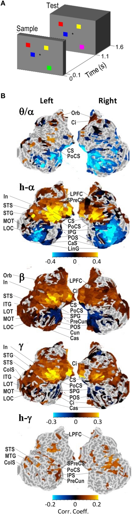
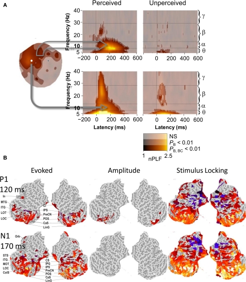
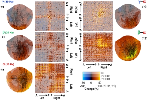
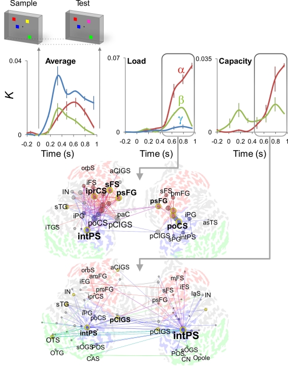
Alpha-frequency band (8-14 Hz) oscillations are among the most salient phenomena in human electroencephalography (EEG) recordings and yet their functional roles have remained unclear. Much of research on alpha oscillations in human EEG has focused on peri-stimulus amplitude dynamics, which phenomenologically support an idea of alpha oscillations being negatively correlated with local cortical excitability and having a role in the suppression of task-irrelevant neuronal processing. This kind of an inhibitory role for alpha oscillations is also supported by several functional magnetic resonance imaging and trans-cranial magnetic stimulation studies. Nevertheless, investigations of local and inter-areal alpha phase dynamics suggest that the alpha-frequency band rhythmicity may play a role also in active task-relevant neuronal processing. These data imply that inter-areal alpha phase synchronization could support attentional, executive, and contextual functions. In this review, we outline evidence supporting different views on the roles of alpha oscillations in cortical networks and unresolved issues that should be addressed to resolve or reconcile these apparently contrasting hypotheses.

Keywords: alpha; amplitude; electroencephalography; magnetoencephalography; phase; source modeling; synchrony.


Figures

Figure 1
Figure 1
Alpha-, beta-, and gamma-band amplitudes are positively correlated with visual working memory (VWM) load in fronto-parietal cortical regions during the retention period. (A) A schematic representation of the VWM paradigm in this study. The subjects sustained a memory of the Sample stimulus and indicated with a force-choice response whether the Test stimulus was identical to the Sample or not. The stimuli contained one to six colored squares in random locations and in the 50% of Test stimuli, the color of one square was different from the color in the Sample stimulus. (B) Cortical localization of the memory-load dependence of oscillation amplitudes during the memory retention period (0.4–1 s). The complete cortical surfaces are flattened and oriented so that the anterior–posterior axis corresponds to up–down direction, respectively. The color values indicate the correlation coefficients obtained across subjects of the oscillation amplitude with the number of objects in the stimuli. The correlation values are shown for cortical regions that were significantly correlated with memory-load. Adapted from Palva et al. (2011).
Figure 2
Figure 2
Post-stimulus phase dynamics of ongoing brain activity in frontal brain regions play a role in perception and working memory encoding. (A) Phase-locking of ongoing alpha, but not of theta or beta oscillations to threshold-level somatosensory stimuli dissociates conscious detection from unconscious stimulus processing. Cortically widespread phase-locking in the alpha-band was observed for perceived stimuli but not for the unperceived stimuli both in signals from a large set of MEG gradiometers over frontal regions (upper panel) as well as in signals from a single sensor over the putative contralateral primary somatosensory region (lower panel). Adapted from Palva et al. (2005b). (B) Cortical localization of evoked responses, positive oscillation amplitude modulations, and phase-locking to stimulus onset to Sample VWM stimuli (see Figure 1) of broadband (1–45 Hz) ongoing brain activity at the latencies of the P1 and N1 components of the visual evoked response. Unlike evoked responses (left panel), phase-locking (right panel) is observed in widespread and task-relevant frontal cortical regions. Conversely, enhanced oscillation amplitudes (middle panel), which are indicative of true stimulus-evoked additive components, are observed only in early visual regions. Color values indicate the response strength in significantly activated cortical regions. Adapted from Palva et al. (2011).
Figure 3
Figure 3
Long-range phase synchronization in the alpha- and to a lesser extent in the beta- and gamma-frequency bands is enhanced during the performance of a continuous working memory task (left panels) in planar MEG gradiometer data. Concurrent cross-frequency phase synchrony between the alpha- and beta-/gamma-frequency bands was also robustly enhanced (right panels). The connectivity matrices indicate the strength of synchronization relative to a rest condition in each MEG gradiometer pair (A, anterior; P, posterior). Adapted from Palva et al. (2005a).
Figure 4
Figure 4
Single-trial source modeling of M/EEG data reveals dynamic phase synchronization during the VWM retention period of a parametric VWM task. Cortical inter-areal phase synchrony, as indexed by connection density K, was robust in alpha (red), beta (green), and gamma (blue) frequency bands throughout the working memory retention period (0.4–1 s) in data averaged across memory-loads from one to six objects (Average, upper panels). During the retention period, these inter-areal interactions were positively correlated with the memory-load (Load, upper panels) in fronto-parietal networks (middle panel, red, frontal; blue, parietal; green, occipital and occipito-temporal; the graph's vertices are brain areas and the edges indicate memory-load dependent inter-areal synchronization in the alpha-band). A plateau in the memory-load dependence of a subset of interactions was positively correlated with the individual subjects’ behavioral working memory capacity (Capacity, upper panels) and networks where the intra-parietal sulcus (intPS) was the most prominent hub (lower panel, alpha-band synchronization correlated with memory capacity). Adapted from Palva et al. (2010).
Figure 5
Figure 5
Local oscillation amplitudes may be suppressed concurrently with strengthened inter-areal synchronization in VWM task retention period (see Figures 1 and 4). The amplitudes (left panel) and networks of inter-areal phase synchronization (right panel) in the alpha-, beta-, and gamma-bands are displayed on flattened cortical surfaces. The amplitude color values (left panel) indicate the mean relative oscillation amplitude change from the baseline level and are shown for significantly modulated regions. The colored edges (right panel) indicate the temporally and spectrally most stable pairs of cortical areas between which the synchronization was strengthened above the baseline level within the alpha-, beta-, and gamma-bands during the VWM retention period. The edge colors indicate the cortical source/target region so that green denotes occipital and occipito-temporal regions, blue the parietal regions, and red the frontal regions. Adapted from Palva et al. (2010, 2011).
Figure 6
Figure 6
The phase of infra-slow (0.01–0.1 Hz) fluctuations is correlated similarly with both the amplitudes of fast (1–40 Hz) oscillations and the detection of threshold-level somatosensory stimuli. This suggests that the phase of infra-slow oscillations reflect global excitability modulations. Adapted from Monto et al. (2008).
Figure 7
Figure 7
The networks of inter-areal synchronization in the alpha and beta bands are more clustered and small-world like than those in the delta- and gamma-frequency bands. C/CR indicates the relative clustering coefficient of the networks in these four frequency bands, whereas σ denotes their small-worldness. Modified from Palva et al. (2010b).

References

    1. Andersen E. A. S. (1968). Physiological Basis of the Alpha Rhythm New York, NY: Appleton-Century-Crofts
    1. Aron A. R., Robbins T. W., Poldrack R. A. (2004). Inhibition and the right inferior frontal cortex. Trends Cogn. Sci. (Regul. Ed.) 8, 170–177 10.1016/j.tics.2004.02.010 - DOI - PubMed
    1. Bar M., Kassam K. S., Ghuman A. S., Boshyan J., Schmid A. M., Dale A. M., Hamalainen M. S., Marinkovic K., Schacter D. L., Rosen B. R., Halgren E. (2006). Top-down facilitation of visual recognition. Proc. Natl. Acad. Sci. U.S.A. 103, 449–454 10.1073/pnas.0507062103 - DOI - PMC - PubMed
    1. Barry R. J., Rushby J. A., Johnstone S. J., Clarke A. R., Croft R. J., Lawrence C. A. (2004). Event-related potentials in the auditory oddball as a function of EEG alpha phase at stimulus onset. Clin. Neurophysiol. 115, 2593–2601 10.1016/S1388-2457(03)00343-2 - DOI - PubMed
    1. Bassett D. S., Bullmore E. (2006). Small-world brain networks. Neuroscientist 12, 512–523 10.1177/1073858406293182 - DOI - PubMed

LinkOut - more resources