. 2011 May;1(3):122-126.
doi: 10.4161/bioa.1.3.17313.

Oskar-induced endocytic activation and actin remodeling for anchorage of the Drosophila germ plasm

Affiliations

Oskar-induced endocytic activation and actin remodeling for anchorage of the Drosophila germ plasm

Tsubasa Tanaka et al. Bioarchitecture. 2011 May.

Abstract

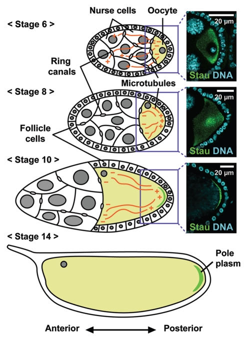
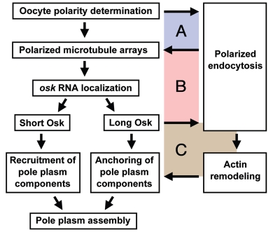
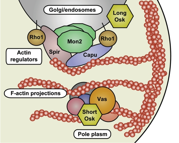
In many animals, germ-cell fate is specified by inheritance of the germ plasm, which is enriched in maternal RNAs and proteins. Assembly of the Drosophila germ (pole) plasm begins with the localization and translation of oskar (osk) RNA at the oocyte posterior pole. osk RNA produces two isoforms, long and short Osk. Short Osk recruits other pole plasm components, and long Osk restricts them to the oocyte cortex. Although molecular functions of long Osk remain mysterious, it is known to be involved in endocytic activation and actin cytoskeletal remodeling. We identified several vesicular trafficking machinery components that act downstream of long Osk in pole plasm assembly. These included the Rab5 effector protein Rabenosyn-5 (Rbsn-5) and the Golgi/endosomal protein Mon2, both of which were crucial for Osk-induced actin remodeling and the anchoring of pole plasm components. We propose that, in response to long Osk, the Rab5/Rbsn-5-dependent endocytic pathway promotes the formation of specialized vesicles, and Mon2 acts on these vesicles as a scaffold to instruct actin nucleators like Cappuccino and Spire to remodel the actin cytoskeleton, which anchors pole plasm components to the cortex. This mechanism may be applicable to the asymmetric localization of macromolecular structures such as protein-RNA complexes in other systems.


Figures

Figure 1
Figure 1
Developmental stages of Drosophila oogenesis. Schematic representations of Drosophila egg chambers. Each egg chamber consists of a single oocyte and 15 nurse cells, which are surrounded by a monolayer of somatic follicle cells. Pole plasm components are synthesized in the nurse cells and transported to the oocyte through the ring canal along the microtubule cytoskeleton, which extends its plus ends into the nurse cells until stage 6. The reorganization of oocyte polarity during stage 6–7 induces a microtubule rearrangement, resulting in polarized microtubule arrays with minus ends along the lateral and anterior cortex and plus ends at the posterior. The polarized microtubule arrays direct the posterior localization of osk RNA, which is subsequently translated into proteins that recruit and anchor pole plasm components to the posterior cortex. Images of osk RNP and nuclei stained with Staufen (Stau) and DAPI, respectively, are shown at right.
Figure 2
Figure 2
Multiple interdependent relationships between pole plasm assembly and endocytosis. The polarized microtubule arrays that are induced by oocyte polarization are required for the initial activation of endocytosis at the posterior, while the increased endocytosis at the oocyte posterior maintains the plus-end targeting of microtubules to the posterior. Thus, the microtubule array polarization and local endocytic activation are interdependent (A). Polarized arrays of microtubules direct the posterior localization of osk RNA, resulting in local synthesis of the two Osk isoforms. Short Osk recruits downstream pole plasm components to the posterior, while long Osk stimulates endocytosis, thereby generating a second positive feedback loop (B). Increased endocytosis promotes the rearrangement of cortical F-actin, resulting in the formation of long F-actin projections, which anchor pole plasm components at the cortex. Because pole plasm anchoring is likely to increase the posterior localization of osk RNA, long Osk-induced endocytosis and actin remodeling form a third positive feedback loop (C).
Figure 3
Figure 3
A model for pole plasm anchoring to the posterior cortex of the Drosophila oocyte. At the oocyte posterior, long Osk stimulates endocytosis, producing specialized vesicles that orchestrate actin remodeling. On these vesicles, Mon2 acts as a platform for actin regulators including Capu, Spir and Rho1, to promote the formation of the long F-actin projections that are crucial for anchoring the pole plasm.

Similar articles

Cited by

References

    1. Spradling AC. Development genetics of oogenesis. In: Bate M, Martinez-Arias A, editors. The Development of Drosophila melanogaster. Cold Spring Harbor, NY: Cold Spring Harbor Laboratory Press; 1993. pp. 1–70.
    1. Steinhauer J, Kalderon D. Microtubule polarity and axis formation in the Drosophila oocyte. Dev Dyn. 2006;235:1455–1468. - PubMed
    1. Clark I, Giniger E, Ruohola-Baker H, Jan LY, Jan YN. Transient posterior localization of a kinesin fusion protein reflects anteroposterior polarity of the Drosophila oocyte. Curr Biol. 1994;4:289–300. - PubMed
    1. Palacios IM, St. Johnston D. Kinesin light chain-independent function of the Kinesin heavy chain in cytoplasmic streaming and posterior localisation in the Drosophila oocyte. Development. 2002;129:5473–5485. - PubMed
    1. Brendza RP, Serbus LR, Duffy JB, Saxton WM. A function for kinesin I in the posterior transport of oskar mRNA and Staufen protein. Science. 2000;289:2120–2122. - PMC - PubMed

LinkOut - more resources