. 2011 Oct;346(1):99-109.
doi: 10.1007/s00441-011-1231-5. Epub 2011 Sep 16.

In situ proliferation and differentiation of macrophages in dental pulp

Affiliations

In situ proliferation and differentiation of macrophages in dental pulp

Yukikatsu Iwasaki et al. Cell Tissue Res. 2011 Oct.

Abstract

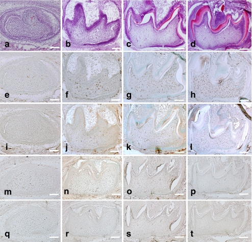
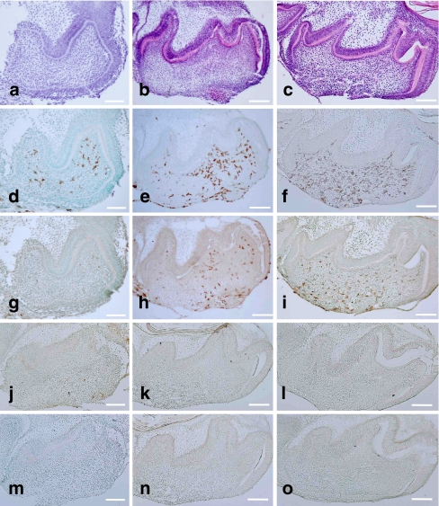
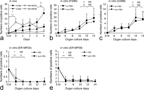
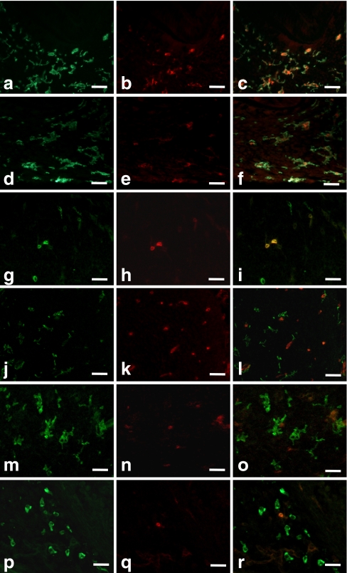
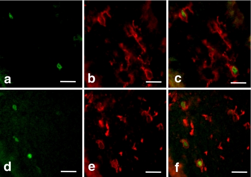
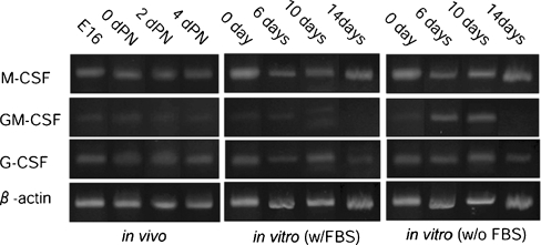
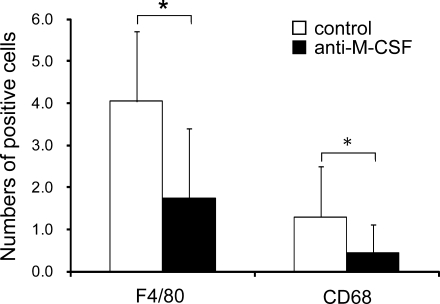
The presence of macrophages in dental pulp is well known. However, whether these macrophages proliferate and differentiate in the dental pulp in situ, or whether they constantly migrate from the blood stream into the dental pulp remains unknown. We have examined and compared the development of dental pulp macrophages in an organ culture system with in vivo tooth organs to clarify the developmental mechanism of these macrophages. The first mandibular molar tooth organs from ICR mice aged between 16 days of gestation (E16) to 5 days postnatally were used for in vivo experiments. Those from E16 were cultured for up to 14 days with or without 10% fetal bovine serum. Dental pulp tissues were analyzed with immunohistochemistry to detect the macrophages and with reverse transcription and the polymerase chain reaction (RT-PCR) for the detection of factors related to macrophage development. The growth curves for the in vivo and in vitro cultured cells revealed similar numbers of F4/80-positive macrophages in the dental pulp. RT-PCR analysis indicated the constant expression of myeloid colony-stimulating factor (M-CSF) in both in-vivo- and in-vitro-cultured dental pulp tissues. Anti-M-CSF antibodies significantly inhibited the increase in the number of macrophages in the dental pulp. These results suggest that (1) most of the dental pulp macrophages proliferate and differentiate in the dental pulp without a supply of precursor cells from the blood stream, (2) M-CSF might be a candidate molecule for dental pulp macrophage development, and (3) serum factors might not directly affect the development of macrophages.


Figures

Fig. 1
Fig. 1
In vivo development of mouse mandibular first molars at E16 (a, e, i, m, q), 0dPN (b, f, j, n, r), 3dPN (c, g, k, o, s), and 5dPN (d, h, l, p, t). Hematoxylin and eosin (H-E) staining indicated the development of tooth organs (a–d). Immunohistochemical detection of F4/80-positive cells (e–h), CD68-positive cells (i–l), ER-MP20-positive cells (m–p), and ER-MP58-positive cells (q–t) indicated an increase in the number of macrophages with tooth organ development. Bars 100 μm
Fig. 2
Fig. 2
Development of E16 mouse mandibular first molars in organ culture under serum-supplemented conditions for 6 days (a, d, g, j, m), 10 days (b, e, h, k, n), and 14 days (c, f, i, l, o). H-E staining indicated the development of tooth organs (a–c). Immunohistochemical detection of F4/80-positive cells (d–f), CD68-positive cells (g–i), ER-MP20-positive cells (j–l), and ER-MP58-positive cells (m–o) indicated an increase in the number of macrophages with tooth organ development. Bar 100 μm
Fig. 3
Fig. 3
Numbers of F4/80- , CD68- , ER-MP20-, and ER-MP58-positive cells in dental pulp with development. Data are representative of means±SD; P values of <0.01 were considered to be significant. a In vivo development of F4/80-, CD68-, ER-MP20-, and ER-MP58-positive cells. b Development of F4/80-positive cells in organ culture with or without fetal bovine serum (w/FBS, w/o FBS). c Development of CD68-positive cells in organ culture with or without fetal bovine serum (w/FBS, w/o FBS). d Development of ER-MP20-positive cells in organ culture with or without fetal bovine serum (w/FBS, w/o FBS). e Development of ER-MP58-positive cells in organ culture with or without fetal bovine serum (w/FBS, w/o FBS). *P<0.01, †: in vivo (F4/80), in vitro (w/FBS), ‡: in vivo (CD68), in vitro (w/o FBS), *: in vivo (ER-MP20), in vitro (w/FBS), **: in vivo (ER-MP58), in vitro (w/o FBS); NS not significant
Fig. 4
Fig. 4
Double-immunostaining of dental pulp macrophages with anti-F4/80 (a, d) and anti-CD68 (b, e) antibodies in 4dPN (a–c) and 14-day-cultured tooth organs (d–f). All CD68-positive cells were F4/80-positive (c, f). Double-immunostaining of dental pulp macrophages with anti-F4/80 (g, j, m, p) and anti-ER-MP20 (h, k, n, q) antibodies in E16 (g–i), 3dPN (j–l), 8-day (m–o), and 14-day-cultured tooth organs (p–r). Bar 25 μm
Fig. 5
Fig. 5
Double-immunostaining of dental pulp macrophages with anti-PCNA (a, d) and anti-F4/80 (b, e) antibodies in 4dPN (a–c) and 8-day-cultured tooth organs (d–f). All PCNA-positive cells were F4/80-positive (c, f). Bar 25 μm
Fig. 6
Fig. 6
RT-PCR analysis of the expression of M-CSF, GM-CSF, G-CSF, and β-actin in dental pulp of in vivo and organ culture with or without fetal bovine serum (w/FBS, w/oFBS)
Fig. 7
Fig. 7
Effects of the M-CSF neutralizing antibody on the development of macrophages in dental pulp. The number of F4/80- and CD68-positive cells was significantly decreased by the addition of the antibody to the medium. Data are representative of means±SD. *P values of <0.01 were considered to be significant

References

    1. Chan J, Leenen PJ, Bertoncello I, Nishikawa SI, Hamilton JA. Macrophage lineage cells in inflammation: characterization by colony-stimulating factor-1 (CSF-1) receptor (c-Fms), ER-MP58, and ER-MP20 (Ly-6C) expression. Blood. 1998;92:1423–1431. - PubMed
    1. Daems WT, Brederoo P. The fine structure and peroxidase activity of resident and exudate peritoneal macrophages in the guinea pig. Adv Exp Med Biol. 1972;15:19–31.
    1. Daems WT, Brederoo P. Electron microscopical studies on the structure, phagocytic properties, and peroxidatic activity of resident and exudate peritoneal macrophages in guinea pigs. Z Zellforsch Mikrosk Anat. 1973;144:247–297. doi: 10.1007/BF00307305. - DOI - PubMed
    1. Daems WT, van der Rhee HJ. Peroxidase and catalase in monocytes, macrophages, epithelioid cells and giant cells of the rat. In: van Furth R, editor. Mononuclear phagocytes, functional aspects. The Hague: Nijhoff; 1980. pp. 43–60.
    1. Daems WT, Koerten HK, Soranzo MR. Differences between monocyte-derived and tissue macrophages. Adv Exp Med Biol. 1976;73:27–40. - PubMed

Publication types

Substances

LinkOut - more resources