. 2012 Oct;33(10):2499-508.
doi: 10.1002/hbm.21380. Epub 2011 Sep 16.

Modulating cortico-striatal and thalamo-cortical functional connectivity with transcranial direct current stimulation

Affiliations

Modulating cortico-striatal and thalamo-cortical functional connectivity with transcranial direct current stimulation

Rafael Polanía et al. Hum Brain Mapp. 2012 Oct.

Abstract

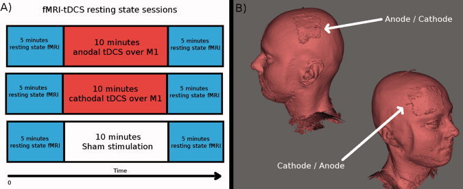
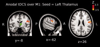
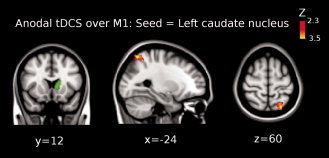
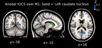
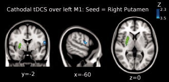
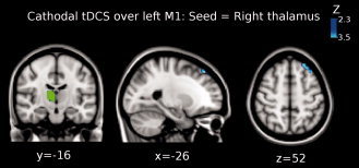
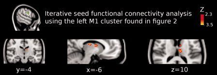
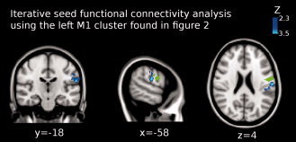
Transcranial direct current stimulation (tDCS) is a noninvasive brain stimulation technique that has been shown to alter cortical excitability and activity via application of weak direct currents. Beyond intracortical effects, functional imaging as well as behavioral studies are suggesting additional tDCS-driven alterations of subcortical areas, however, direct evidence for such effects is scarce. We aimed to investigate the impact of tDCS on cortico-subcortical functional networks by seed functional connectivity analysis of different striatal and thalamic regions to prove tDCS-induced alterations of the cortico-striato-thalamic circuit. fMRI resting state data sets were acquired immediately before and after 10 min of bipolar tDCS during rest, with the anode/cathode placed over the left primary motor cortex (M1) and the cathode/anode over the contralateral frontopolar cortex. To control for possible placebo effects, an additional sham stimulation session was carried out. Functional coupling between the left thalamus and the ipsilateral primary motor cortex (M1) significantly increased following anodal stimulation over M1. Additionally, functional connectivity between the left caudate nucleus and parietal association cortices was significantly strengthened. In contrast, cathodal tDCS over M1 decreased functional coupling between left M1 and contralateral putamen. In summary, in this study, we show for the first time that tDCS modulates functional connectivity of cortico-striatal and thalamo-cortical circuits. Here we highlight that anodal tDCS over M1 is capable of modulating elements of the cortico-striato-thalamo-cortical functional motor circuit.


Figures

Figure 1
Figure 1
(A) Shown is the experimental procedure. First 5 min resting state fMRI was acquired. Then 10 min tDCS was applied inside the MR scanner. Immediately afterwards, a new 5 min resting state fMRI was acquired. Each subject underwent three sessions: anodal tDCS over M1 (combined with cathodal tDCS over the contralateral frontopolar cortex), cathodal tDCS over M1 (combined with anodal tDCS over the contralateral frontopolar cortex) and sham stimulation. The order of sessions was interindividually randomized and the single sessions were separated by at least 8 days from each other. (B) A 3‐dimensional reconstruction of the T1 image from one of the subjects is shown to illustrate the location of the electrodes. For anodal stimulation over M1 the anode was placed over the left M1 and the cathode over the contralateral frontopolar cortex. For cathodal stimulation over M1 the current flux was reversed. tDCS was applied inside the MR scanner, but not during EPI fMRI acquisition.
Figure 2
Figure 2
Shown is the result of the third‐level mixed effects (ME) analysis (see Methods) when using the left thalamus as seed (green colored region in the brain) for the functional connectivity analysis when anodal tDCS was applied over M1. Functional connectivity significantly increased between left thalamus and left precentral gyrus (Brodmann area (BA) 4; peak Z‐value = 3.03, MNI x = −62, y = −8, z = 26; cluster size = 206 voxels). Images are displayed according to radiological convention (left is right).
Figure 3
Figure 3
Shown is the result of the third‐level mixed effects (ME) analysis (see Methods) when using the left caudate nucleus as seed (green colored region in the brain) for the functional connectivity analysis when anodal tDCS was applied over M1. Functional connectivity significantly increased between left caudate nucleus and left superior parietal lobule (Brodmann area 7; peak Z‐value = 3.12, MNI x = −24, y = −62, z = 60; cluster size = 228 voxels). Images are displayed according to radiological convention (left is right).
Figure 4
Figure 4
Shown is the result of the third‐level mixed effects (ME) analysis (see Methods) when using the left caudate nucleus as seed (green colored region in the brain) for the functional connectivity analysis when anodal tDCS was applied over M1. Functional connectivity significantly decreased between left caudate nucleus and posterior cingulate cortex (PCC) (peak Z‐value = 2.91, MNI x = −10, y = −58, z = 16; cluster size = 223 voxels). Images are displayed according to radiological convention (left is right).
Figure 5
Figure 5
Shown is the result of the third‐level mixed effects (ME) analysis (see Methods) when using the right putamen (green colored region in the brain) for the functional connectivity analysis when cathodal tDCS was applied over M1. Functional connectivity significantly decreased between right putamen and left precentral gyrus (Brodmann area 4; peak Z‐value = 3.4, MNI x = −60, y = −2, z = 14; cluster size = 156 voxels). Images are displayed according to radiological convention (left is right).
Figure 6
Figure 6
Shown is the result of the third‐level mixed effects (ME) analysis (see Methods) when using the right thalamus as seed (green colored region in the brain) for the functional connectivity analysis when cathodal tDCS was applied over M1. Functional connectivity significantly decreased between right thalamus and left superior frontal gyrus (peak Z‐value = 3.3, MNI x = −26, y = 36, z = 52; cluster size = 206 voxels). Images are displayed according to radiological convention (left is right).
Figure 7
Figure 7
Shown is the result of the iterative functional connectivity analysis using the left M1 cluster as seed (green colored region). In Figure 2, it is shown that this cluster enhanced functional coupling with the left thalamus (see figure 2) when anodal tDCS was applied over M1. Here, it is shown that functional connectivity significantly increased between left thalamus and left precentral gyrus (peak Z‐value = 2.9; MNI x = −10, y = −16, z = 14; cluster size = 112 voxels). We found a second cluster that significantly increased functional coupling with the left M1, which represents the left thalamus and left caudate nucleus (peak Z‐value = 3.2; MNI x = −6, y = −4, z = 10; cluster size = 150 voxels). Images are displayed according to radiological convention (left is right).
Figure 8
Figure 8
Shown is the result of the iterative functional connectivity analysis using the left M1 cluster as seed (green colored region). In Figure 2, it is shown that this cluster enhanced the functional coupling with the left thalamus (see Fig. 2) when anodal tDCS was applied over M1. Functional connectivity significantly decreased between left M1 and left primary somatosensory cortex (Brodmann area 2; peak Z‐value = 3.51, MNI x = −58, y = −18, z = 3; cluster size = 351 voxels). Images are displayed according to radiological convention (left is right).

References

    1. Amunts K, Jancke L, Mohlberg H, Steinmetz H, Zilles K ( 2000): Interhemispheric asymmetry of the human motor cortex related to handedness and gender. Neuropsychologia 38: 304–312. - PubMed
    1. Antal A, Terney D, Kuhnl S, Paulus W ( 2010): Anodal transcranial direct current stimulation of the motor cortex ameliorates chronic pain and reduces short intracortical inhibition. J Pain Symptom Manage 39: 890–903. - PubMed
    1. Bachmann CG, Muschinsky S, Nitsche MA, Rolke R, Magerl W, Treede RD, Paulus W, Happe S ( 2010): Transcranial direct current stimulation of the motor cortex induces distinct changes in thermal and mechanical sensory percepts. Clin Neurophysiol 121: 2083–2089. - PubMed
    1. Beckmann CF, DeLuca M, Devlin JT, Smith SM ( 2005): Investigations into resting‐state connectivity using independent component analysis. Philos Trans R Soc Lond B Biol Sci 360: 1001–1013. - PMC - PubMed
    1. Benninger DH, Lomarev M, Lopez G, Wassermann EM, Li X, Considine E, Hallett M ( 2010): Transcranial direct current stimulation for the treatment of Parkinson's disease. J Neurol Neurosurg Psychiatry 81: 1105–1111. - PMC - PubMed

Publication types