

Cloudy and starry milia-like cysts: how well do they distinguish seborrheic keratoses from malignant melanomas?
- PMID: 21923811
- PMCID: PMC3212814
- DOI: 10.1111/j.1468-3083.2010.03920.x
Cloudy and starry milia-like cysts: how well do they distinguish seborrheic keratoses from malignant melanomas?
Abstract
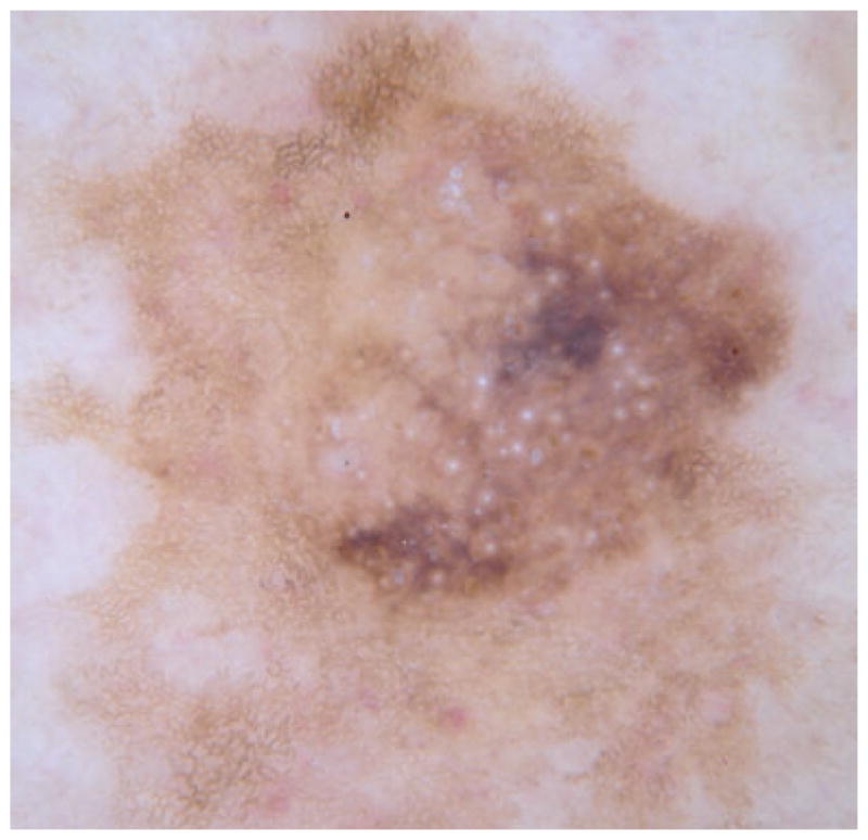
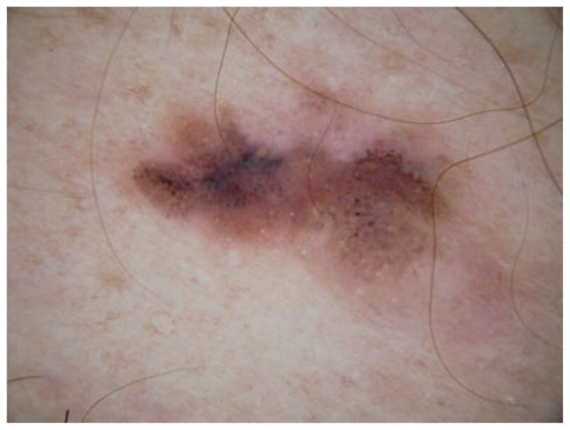
Background: Seborrheic keratoses are the most common skin lesions known to contain small white or yellow structures called milia-like cysts (MLCs). Varied appearances can sometimes make it difficult to differentiate benign lesions from malignant lesions such as melanoma, the deadliest form of skin cancer found in humans.
Objective: The purpose of this study was to determine the statistical occurrence of MLCs in benign vs. malignant lesions.
Methods: A medical student with 10 months experience in examining approximately 1000 dermoscopy images and a dermoscopy-naïve observer analysed contact non-polarized dermoscopy images of 221 malignant melanomas and 175 seborrheic keratoses for presence of MLCs.
Results: The observers found two different types of MLCs present: large ones described as cloudy and smaller ones described as starry. Starry MLCs were found to be prevalent in both seborrheic keratoses and melanomas. Cloudy MLCs, however, were found to have 99.1% specificity for seborrheic keratoses among this group of seborrheic keratoses and melanomas.
Conclusion: Cloudy MLCs can be a useful tool for differentiating between seborrheic keratoses and melanomas.
© 2010 The Authors. Journal of the European Academy of Dermatology and Venereology © 2010 European Academy of Dermatology and Venereology.
Conflict of interest statement
None reported.
Figures


References
-
- Argenziano G, Soyer HP, Chimenti S, et al. Dermoscopy of pigmented skin lesions: results of a consensus meeting via the internet. J Am Acad Dermatol. 2003;48:679–693. - PubMed
-
- Changchien L, Dusza SW, Agero AL, et al. 1. Age- and site-specific variation in the dermoscopic patterns of congenital melanocytic nevi: an aid to accurate classification and assessment of melanocytic nevi. 1. Arch Dermatol. 2007;143:1007–1014. - PubMed
-
- Berk DR, Bayliss SJ. Milia: a review and classification. J Am Acad Dermatol. 2008;59:1050–1063. - PubMed
-
- Menzies SW, Kreusch J, Byth K, et al. Dermoscopic evaluation of amelanotic and hypomelanotic melanoma. Arch Dermatol. 2008;144:1120–1127. - PubMed
-
- Burroni M, Nami N, Rubegni P. Like milia-like cysts. Skin Res Technol. 2009;15:250–251. - PubMed
Publication types
MeSH terms
Grants and funding
LinkOut - more resources
Full Text Sources
Medical
