. 2011 Sep 16:12:41.
doi: 10.1186/1471-2199-12-41.

LIM kinase 1 - dependent cofilin 1 pathway and actin dynamics mediate nuclear retinoid receptor function in T lymphocytes

Affiliations

LIM kinase 1 - dependent cofilin 1 pathway and actin dynamics mediate nuclear retinoid receptor function in T lymphocytes

Mohammad Ishaq et al. BMC Mol Biol. .

Erratum in

  • BMC Mol Biol. 2012;13:14. Aguilera-Gutierrez, Angelica [added]

Abstract

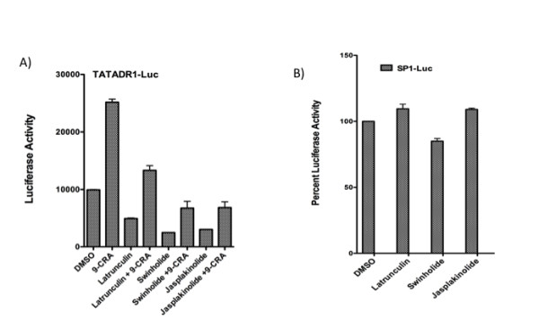
Background: It is known that retinoid receptor function is attenuated during T cell activation, a phenomenon that involves actin remodeling, suggesting that actin modification may play a role in such inhibition. Here we have investigated the role of actin dynamics and the effect of actin cytoskeleton modifying agents on retinoid receptor-mediated transactivation.

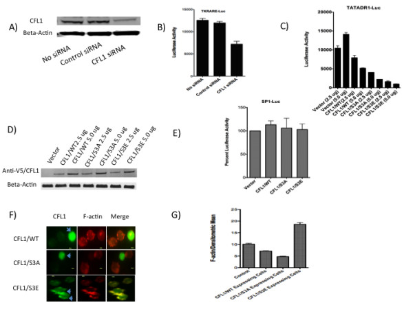
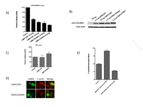
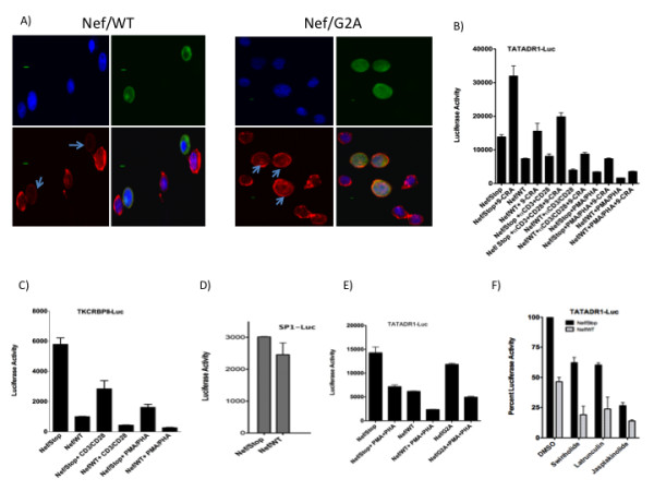
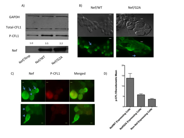
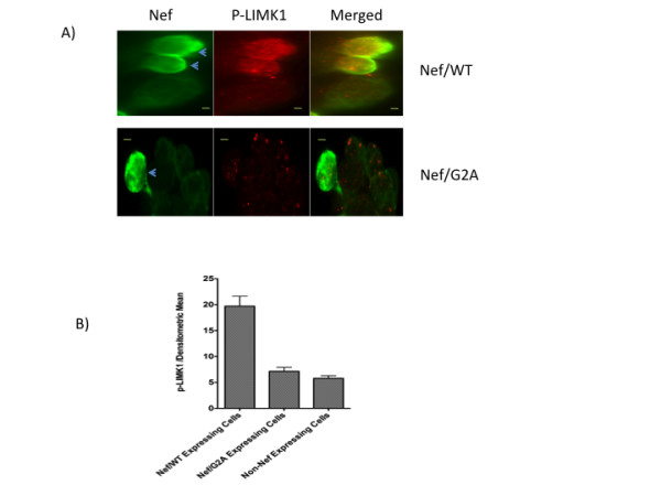
Results: Agents that disturb the F-actin assembly or disassembly attenuated receptor-mediated transcription indicating that actin cytoskeletal homeostasis is important for retinoid receptor function. Overexpression or siRNA-induced knockdown of cofilin-1 (CFL1), a key regulator of F-actin assembly, induced the loss of receptor function. In addition, expression of either constitutively active or inactive/dominant-negative mutants of CFL1or CFL1 kinase LIMK1 induced loss of receptor function suggesting a critical role of the LIMK1-mediated CFL1 pathway in receptor-dependent transcription. Further evidence of the role of LMK1/CFL1-mediated actin dynamics, was provided by studying the effect of Nef, an actin modifying HIV-1 protein, on receptor function. Expression of Nef induced phosphorylation of CFL1 at serine 3 and LIMK1 at threonine 508, inhibited retinoid-receptor mediated reporter activity, and the expression of a number of genes that contain retinoid receptor binding sites in their promoters. The results suggest that the Nef-mediated inhibition of receptor function encompasses deregulation of actin filament dynamics by LIMK1 activation and phosphorylation of CFL1.

Conclusion: We have identified a critical role of LIMK1-mediated CFL1 pathway and actin dynamics in modulating retinoid receptor mediated function and shown that LIMK1-mediated phosphocycling of CFL1 plays a crucial role in maintaining actin homeostasis and receptor activity. We suggest that T cell activation-induced repression of nuclear receptor-dependent transactivation is in part through the modification of actin dynamics.


Figures

Figure 1
Figure 1
F-actin-modifying chemicals attenuate retinoid receptor-mediated transcription. Jurkat cells were transfected with 5.0 ug TATADR1-Luc (A) or SP1-Luc plasmids (B). After 24 h, TATADR1-Luc transfected cells were treated for 16 h with 9-cis retinoic acid (1.0 uM) or vehicle in the presence of DMSO, or actin inhibitors latrunculin A (50 nM), swinholide A (2.5 nM) or jasplakinolide (50 nM). After 24 h, Sp1-Luc transfected cells were treated with DMSO or actin inhibitors for 16 h. Cells were harvested and luciferase activity measured as described in Experimental Procedures.
Figure 2
Figure 2
CFL1 regulates retinoid receptor function. A and B) Jurkat cells were transfected with 2.5 ug of TKRARE-Luc plasmid either in the presence or absence of 2.5 uM control siRNA or CFL1-specific siRNA. Cells were harvested after 36 h and lysates subject to Western blotting using antibodies to CFL1 and beta-actin (A) and luciferase activity measurement (B) as described in Experimental Procedures. C and D) Jurkat cells were transfected with 5.0 ug TATADR1-Luc in the presence of 2.5 ug and 5.0 ug of indicated plasmids. Cells were harvested after 36 h and luciferase activity measured as described in Experimental Procedures (C). Lysates were also subject o Western blotting using antibodies to anti-V5 tag (1:2,000) and beta-actin (1:10,000) (D). E) Jurkat cells were transfected with 5.0 ug of SP1-Luc plasmid in the presence of 5 ug of indicated plasmids. Cells were harvested after 36 h and luciferase activity measured as described in Experimental Procedures. SP1-Luc activity obtained in the presence of vector was normalized to 100%. F and G) Jurkat cells were transfected with CFL1/WT, CFL1/S3A, and CFL1/S3E plasmids and stained with anti-V5 Tag antibodies followed by treatment with F-actin stain as per the instructions of the manufacturer. The cells were visualized using immunofluorescent microscopy (F) and the densitometric mean intensity of F-actin from V5-CFL1 positive cells was quantified and compared to non-V5-CFL1 expressing control cells (G) using AxioVision software from Zeiss Imager D1 fluorescent microscope. Bar, 2 um.
Figure 3
Figure 3
LIMK1 regulates retinoid receptor function. A and B) Jurkat cells were transfected with 5.0 ug TATADR1-Luc in the presence of 2.5 ug and 5.0 ug of indicated plasmids. Cells were harvested after 36 h and luciferase activity measured as described in Experimental Procedures (A). Lysates were also subject o Western blotting using antibodies to anti-V5 tag (1:2,000) and beta-actin (1:10,000) (B). C) Jurkat cells were transfected with 5.0 ug of SP1-Luc plasmid in the presence of 5 ug of indicated plasmids. Cells were harvested after 36 h and luciferase activity measured as described in Experimental Procedures. SP1-Luc activity obtained in the presence of vector was normalized to 100%. D and E) Jurkat cells were transfected with LIMK1/WT, and CFL1/D460A plasmids and stained with anti-V5 Tag antibodies followed by treatment with F-actin stain. The cells were visualized using immunofluorescent microscopy (D) and the densitometric mean intensity of F-actin from V5-LIMK1 positive cells was quantified and compared to non-V5-LIMK1 expressing control cells (E) using AxioVision software from Zeiss Imager D1 fluorescent microscope. Bar, 2 um.
Figure 4
Figure 4
HIV-1 Nef induces loss of F-actin assembly and inhibits retinoid receptor-mediated transcription. A) Jurkat cells were transfected for 36 h with 5.0 ug of Nef/WT or Nef/G2A plasmids using Fugene-HD. The cells were fixed, permeabilized, and blocked with BSA as described in Experimental Procedures. Cells were incubated with anti-Nef anti-serum (1:500 in 5% BSA/PBS) for 1-2 h and labeled with FITC-conjugated secondary antibody. Cells were stained for F-actin using Alexa Fluor 555 Phalloidin and mounted in Prolong Gold Antifade Reagent with DAPI. Confocal images were obtained using an Olympus Fluoview 1000-inverted microscope. Each figure is a quadron for DAPI, Nef, F-actin, and merged images. F-actin stained Nef expressing cells are indicated by arrows. Bar, 2.5 um. B-E) Jurkat (B, E) or PBT (C) cells were transfected with 5.0 ug TATADR1-Luc or TKCRBPII-Luc plasmids respectively in the presence of 2.5 ug of indicated plasmids. After 16 h, cells were treated as indicated and luciferase activity measured 24 h later as described in Materials and Methods. D) Jurkat cells were transfected with 2.5 ug of SP1-Luc in the presence of indicated plasmids and luciferase activity measured 36 h later. F) Jurkat cells were transfected with 5.0 ug TATADR1-Luc in the presence of 2.5 ug of indicated plasmids. After 16 h, cells were treated with indicated compounds and luciferase activity measured 24 h later as described in Experimental Procedures.
Figure 5
Figure 5
Nef induces CFL1 phosphorylation. A) 293 cells were transfected with Nef/Stop, Nef/WT or Nef/G2A plasmids for 36 h. Cell lysates were then subject to Western blotting using antibodies to Nef (1:2,000), total CFL1 (1:1,000), phospho-serine-3-CFL1 (1:2,000), and GAPDH (1:5,000). The intensity of the phospho-serine-3-CFL1 bands was quantified and normalized to Nef/Stop that was assigned a value of 1.0. B and C) 293 cells transfected with either Flag-tagged Nef/WT or Flag-tagged Nef/G2A plasmids were fixed, permeabilized, and blocked with BSA (as described in Experimental Procedures), incubated with anti-Flag (B) or anti-Flag and anti-phospho-serine-3-CFL1 antibodies (C) (1:500 in 5% BSA/PBS) for 2 h, and stained with FITC-conjugated (B) or FITC-conjugated and Alex-Fluor-555 labeled secondary antibodies (C). The cells were mounted in Prolong Gold Antifade Reagent before immunofluorescence microscopy. Fig 5B also shows the phase contrast image of 293 cells expressing Flag-tagged Nef/WT exhibiting "rounding off" phenotype unlike cells expressing Flag-tagged Nef/G2A. The arrows in Fig. 5B identify non-Nef expressing cells. The panels in Fig 5C are for Nef, p-CFL1, and merged images. The arrows in Fig. 5C identify Nef expressing cells. Bar, 5 um. D) The densitometric mean intensity of p-CFL1 stained cells was quantified from Nef/WT and Nef/G2A transfected cells and compared to non-Nef expressing cells.
Figure 6
Figure 6
Nef induces LIMK1 activation. 293 cells were transfected with either Flag-tagged Nef/WT (A) or Flag-tagged Nef/G2A (B) plasmids. After 36 h cells were fixed, permeabilized, and blocked with BSA as described in Experimental Procedures. Cells were incubated with anti-Flag tag (1:500 in 5% BSA/PBS) and phospho-specific LIMK1 (1:250 in 5% BSA/PBS) antibodies for 2 h and labeled with FITC-conjugated (Flag) and Alex-Fluor-555 labeled (phospho-CFL1) secondary antibodies. The cells were mounted in Prolong Gold Antifade Reagent before immunofluorescence microscopy. The panels in the figure are for Nef, p-LIMK1, and merged images. Arrows identify Nef expressing cells. Bar, 5 um. B) The densitometric mean intensity of p-LIMK1 stained cells was quantified from Nef/WT and Nef/G2A transfected cells and compared to non-Nef expressing cells.

Similar articles

Cited by

References

    1. Curotto de Lafaille MA, Lafaille JJ. Natural and Adaptive Foxp3+ Regulatory T Cells: More of the Same or a Division of Labor? Immunity. 2009;30:626–635. doi: 10.1016/j.immuni.2009.05.002. - DOI - PubMed
    1. Klemann C, Ravney BJ, Oki S, Yamamura T. Retinoid signals and Th17-mediated pathology. Nihon Rinsho Meneki Gakkai Kaishi. 2009;32:20–28. doi: 10.2177/jsci.32.20. - DOI - PubMed
    1. Mucida D, Park Y, Kim G, Turovskaya O, Scott I, Kronenberg M, Cheroutre H. Reciprocal TH17 and Regulatory T Cell Differentiation Mediated by Retinoic Acid. Science. 2007;317:256–260. doi: 10.1126/science.1145697. - DOI - PubMed
    1. Sun CM, Hall JA, Blank RB, Bouladoux N, Oukka M, Mora JR, Belkaid Y. Small intestine lamina propria dendritic cells promote de novo generation of Foxp3 T reg cells via retinoic acid. J Exp Med. 2007;204:1775–1785. doi: 10.1084/jem.20070602. - DOI - PMC - PubMed
    1. Ishaq M, DeGray G, Natarajan V. Evidence for the involvement of tyrosine kinase ZAP 70 in nuclear retinoid receptor-dependent transactivation in T lymphocytes. J Biol Chem. 2005;280:34152–34158. doi: 10.1074/jbc.M501547200. - DOI - PubMed

Publication types

MeSH terms

Associated data