. 2011 Nov;179(5):2337-45.
doi: 10.1016/j.ajpath.2011.07.047. Epub 2011 Sep 13.

Macrophage-mediated dorsal root ganglion damage precedes altered nerve conduction in SIV-infected macaques

Affiliations

Macrophage-mediated dorsal root ganglion damage precedes altered nerve conduction in SIV-infected macaques

Victoria A Laast et al. Am J Pathol. 2011 Nov.

Abstract

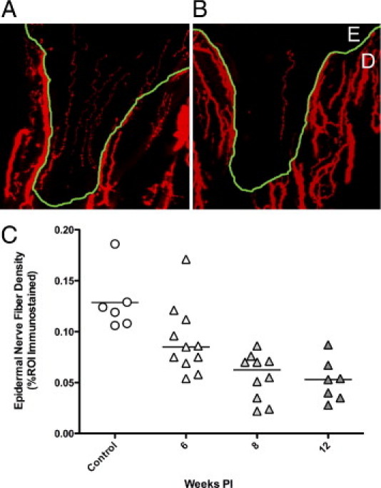
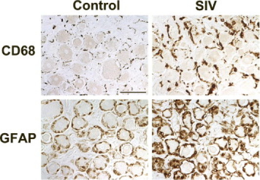
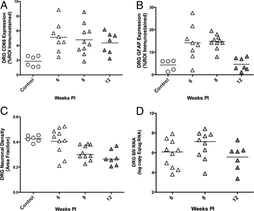
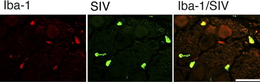
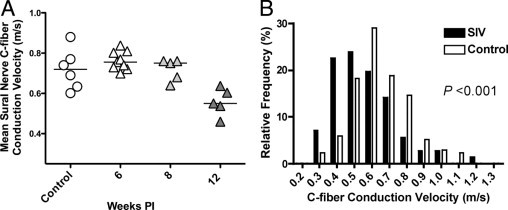
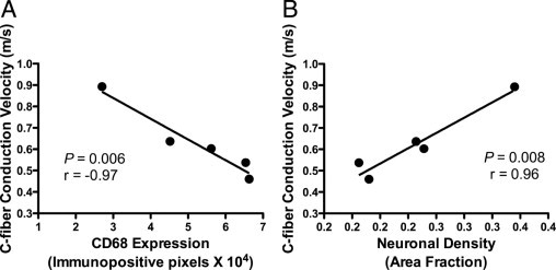
Peripheral neuropathy is the most common neurological complication of HIV-1 infection, affecting over one-third of infected individuals, including those treated with antiretroviral therapy. To study the pathogenesis of HIV-induced peripheral nervous system disease, we established a model in which SIV-infected macaques developed changes closely resembling alterations reported in components of the sensory pathway in HIV-infected individuals. Significant declines in epidermal nerve fiber density developed in SIV-infected macaques, similar to that of HIV-infected individuals with neuropathy. Changes in dorsal root ganglia (DRG) included macrophage infiltration, SIV replication in macrophages, immune activation of satellite cells, and neuronal loss. To determine whether dorsal root ganglion damage was associated with altered nerve function, we measured unmyelinated C-fiber conduction velocities (CV) in nerves of SIV-infected macaques and compared CV changes with DRG alterations. Twelve weeks postinoculation, SIV-infected macaques had significantly lower C-fiber conduction velocity in sural nerves than uninfected animals and the magnitude of conduction velocity decline correlated strongly with extent of DRG macrophage infiltration. Thus, injury to neurons in the DRG-mediated by activated macrophages-preceded altered conduction of unmyelinated nerve fibers in SIV-infected macaques, suggesting that macrophage-mediated DRG damage may be the initiating event in HIV-induced sensory neuropathy.


Figures

Figure 1
Figure 1
Epidermal nerve fiber density is reduced with SIV infection. PGP9.5 immunostained sections of footpad from control (A) and SIV-infected macaques (B) showed a decline in epidermal nerve fiber density. Nerve fibers (red) in skin are present in both epidermis (E), and in dermis (D), with dermal-epidermal junction traced in green. C: Measurement of ENF density in control and SIV-infected macaques demonstrated reduced ENF density in SIV-infected macaques (triangles) compared to uninfected controls (circles), with significant ENF decline beginning 8 weeks p.i., then declining further at the 12-week time point. P < 0.001, analysis of variance). PI, postinoculation; ROI, region of interest.
Figure 2
Figure 2
Increased CD68 and GFAP expression in DRG of SIV-infected macaques. Immunostaining for the macrophage marker CD68 (top panels) and the satellite cell marker GFAP (bottom panels) in the lumbar DRG was increased in SIV-infected macaques versus uninfected control animals. Sections of DRG from control macaques contained scattered CD68-positive resident macrophages diffusely distributed in the perineuronal compartment, whereas SIV-infected macaques had increased immunostaining for CD68 both in cells consistent with infiltrating macrophages and diffusely in activated, endogenous macrophages in DRG. Scale bar = 50 μm.
Figure 3
Figure 3
Alterations in CD68, GFAP, neuronal density, and SIV RNA in DRG over time. A: Scatter plot depicting the amount of CD68 immunostaining for macrophages in the dorsal root ganglia of control macaques (circles) and SIV-infected macaques (triangles) demonstrates a statistically significant increase in macrophage activation and infiltration in the DRG of SIV-infected macaques beginning 6 weeks p.i. and then sustained at comparable levels throughout the course of infection to 12 weeks p.i. (P = 0.008, analysis of variance). B: A similar significant increase in GFAP immunostaining of satellite cells was measured 6 weeks p.i. as well as 8 weeks p.i., before declining to control animal levels 12 weeks. p.i. (P = 0.001, analysis of variance). C: Neuronal area fraction measurements in the dorsal root ganglia of control macaques and SIV-infected macaques demonstrated a statistically significant decrease in neuronal density in the dorsal root ganglia of SIV-infected macaques 12 weeks p.i. as compared to control macaques (P = 0.001, analysis of variance,). D: SIV RNA levels in DRG were stable from 6 to 12 weeks p.i., consistent with sustained CD68 expression over time. Bars represent group means. PI, postinoculation; ROI, region of interest.
Figure 4
Figure 4
SIV infection of macrophages in the DRG. Confocal laser scanning microscopy was performed on sections of dorsal root ganglia from SIV-infected macaques double immunostained for the macrophage marker Iba-1 (red, left) and SIV gp41 (green, middle). A merged image showed colocalization of Iba-1 and SIV gp41 (yellow, right), indicating that macrophages were the predominant SIV-infected cell type in sensory ganglia.
Figure 5
Figure 5
Decreased C-fiber conduction velocity in SIV-infection. A: Mean sural nerve C-fiber conduction velocity for the group of SIV-infected animals evaluated 12 weeks p.i. was significantly slower than for uninfected control animals as well as for the groups of SIV-infected animals evaluated 6 and 8 weeks postinoculation (P = 0.003, analysis of variance). B: To characterize the functional alterations in peripheral nerves that developed with SIV infection, the conduction velocities of C-fibers in sural nerves from SIV-infected monkeys collected 12 weeks p.i. were compared with the conduction velocities in uninfected control animals. The histogram containing the pooled C-fiber CV measurements for SIV-infected (black bars) and control macaques (white bars) showed an increase in the percentage of nerve fibers with slower conduction velocities in SIV-infected macaques 12 weeks p.i. as compared to controls (P < 0.001; Student's t-test). PI, postinoculation.
Figure 6
Figure 6
C-fiber conduction velocity correlated strongly with DRG alterations. Comparing the mean C-fiber conduction velocity of SIV-infected animals 12 weeks p.i with corresponding lumbar DRG alterations demonstrated a strong inverse correlation between slowing of CV in C-fibers and increased macrophage infiltration and activation denoted by levels of CD68 immunostaining within DRG (A, P = 0.006, r = −0.97, Pearson correlation coefficient). The mean C-fiber conduction velocity was also strongly correlated with neuronal density in lumbar DRG (B, P = 0.008, r = 0.96, Pearson correlation coefficient).

References

    1. McArthur J.C., Brew B.J., Nath A. Neurological complications of HIV infection. Lancet Neurol. 2005;4:543–555. - PubMed
    1. Ellis R.J., Rosario D., Clifford D.B., McArthur J.C., Simpson D., Alexander T., Gelman B.B., Vaida F., Collier A., Marra C.M., Ances B., Atkinson J.H., Dworkin R.H., Morgello S., Grant I. Continued high prevalence and adverse clinical impact of human immunodeficiency virus-associated sensory neuropathy in the era of combination antiretroviral therapy: the CHARTER Study. Arch Neurol. 2010;67:552–558. - PMC - PubMed
    1. Cornblath D.R., McArthur J.C. Predominantly sensory neuropathy in patients with AIDS and AIDS-related complex. Neurology. 1988;38:794–796. - PubMed
    1. Pardo C.A., McArthur J.C., Griffin J.W. HIV neuropathy: insights in the pathology of HIV peripheral nerve disease. J Peripher Nerv Syst. 2001;6:21–27. - PubMed
    1. Simpson D.M., Tagliati M. Neurologic manifestations of HIV infection. Ann Intern Med. 1994;121:769–785. - PubMed

Publication types

MeSH terms

Substances