

Increased T wave complexity can indicate subclinical myocardial ischemia in asymptomatic adults
- PMID: 21924433
- PMCID: PMC3200448
- DOI: 10.1016/j.jelectrocard.2011.07.017
Increased T wave complexity can indicate subclinical myocardial ischemia in asymptomatic adults
Abstract
Background: Altered ventricular repolarization and cardiovascular mortality are closely correlated, and recent novel findings show that a distorted T wave loop morphology is also strongly correlated with subsequent onset of myocardial infarction among patients with stable angina. Therefore, we hypothesized that an abnormal T wave complexity ratio (CR) can indicate vulnerability to myocardial ischemia in asymptomatic, apparently healthy adults.
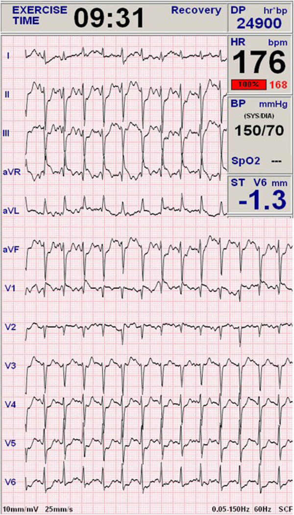
Methods: Healthy firefighters were enrolled in the current investigation where they completed symptom-limited, graded exercise treadmill testing (ETT) and 24-hour Holter electrocardiogram recording. The CR was automatically calculated using principal component analysis of the high-resolution Holter electrocardiogram signal then averaged over 24 hours (CR(24h)). End points were manually analyzed from the ETT; recordings revealing horizontal ST-segment depression (≥ 1 mm) in 2 or more leads for at least 1 minute during the peak of exercise were considered indicators of myocardial ischemia.
Results: One hundred four firefighters (age, 44 ± 8 years; 96% men) completed both ETT and Holter recording. Firefighters with positive end points (n = 34, or 33%) had higher CR(24h) compared with those with negative end points (0.14 ± 0.06 vs 0.09 ± 0.04, P < .01); there were no demographic differences between the 2 groups. After controlling for age, smoking status, hypertension, and obesity, an abnormal CR(24h) (≥ 20%) significantly predicted exercise-induced myocardial ischemia (odds ratio, 4.6; P = .01).
Conclusions: Increased T wave CR(24h) can predict myocardial ischemia in asymptomatic middle-age adults; this suggests that the distorted T wave loop morphology can reflect an altered ventricular repolarization caused by prolonged subclinical myocardial ischemia possibly caused by early coronary artery disease.
Copyright © 2011 Elsevier Inc. All rights reserved.
Figures
References
-
- Pentti MR. Dispersed measures of dispersed repolarization and depolarization: Scalars, vectors, angles, maps, and prospects for improved clinical utility. Journal of Electrocardiology. 2010;43(4):283–287. - PubMed
-
- Priori SG, Mortara DW, Napolitano C, et al. Evaluation of the Spatial Aspects of T-Wave Complexity in the Long-QT Syndrome. Circulation. 1997;96(9):3006–3012. - PubMed
-
- Prineas RJ, Grandits G, Rautaharju PM, Cohen JD, Zhang ZM, Crow RS. Long-term prognostic significance of isolated minor electrocardiographic T-wave abnormalities in middle-aged men free of clinical cardiovascular disease (The Multiple Risk Factor Intervention Trial [MRFIT]) American Journal of Cardiology. 2002;90(12):1391–1395. - PubMed
-
- Okin P, Devereux R, Fabsitz R, Lee E, Galloway J, Howard B. Principal Component Analysis of the T Wave and Prediction of Cardiovascular Mortality in American Indians: The Strong Heart Study. Circulation. 2002;105(6):714–719. - PubMed
Publication types
MeSH terms
Grants and funding
LinkOut - more resources
Full Text Sources
Other Literature Sources
