Review
. 2011:74:13-53.
doi: 10.1016/B978-0-12-387690-4.00002-7.

The itty-bitty time machine genetics of the cyanobacterial circadian clock

Affiliations
Review

The itty-bitty time machine genetics of the cyanobacterial circadian clock

Shannon R Mackey et al. Adv Genet. 2011.

Abstract

The cyanobacterium Synechococcus elongatus PCC 7942 has been used as the prokaryotic model system for the study of circadian rhythms for the past two decades. Its genetic malleability has been instrumental in the discovery of key input, oscillator, and output components and has also provided monumental insights into the mechanism by which proteins function to maintain and dictate 24-h time. In addition, basic research into the prokaryotic system has led to interesting advances in eukaryotic circadian mechanisms. Undoubtedly, continued genetic and mutational analyses of this single-celled cyanobacterium will aid in teasing out the intricacies of the Kai-based circadian clock to advance our understanding of this system as well as other more "complex" systems.


Figures

Figure 1
Figure 1
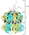
Period-altering amino acid substitutions mapped to structure of a KaiC hexamer. KaiC monomers consist of two tandemly duplicated domains, CI and CII. These monomers oligomerize into hexameric structures with a thin “waist” region connecting the CI and CII regions. At the C-terminus of each KaiC monomer exists an “A-loop,” which determines the steady-state level of KaiC phosphorylation. Amino acid substitutions in the KaiC protein (Ishiura et al., 1998) that result in short-period rhythms (A87V, R215C, and R321Q) are denoted by gray circles, while those substitutions that give rise to period lengths greater than that of wild-type cells (S157C, P236S, R253H, M273I, T409A, and Y442H) are depicted as black circles. Image provided by Yong-Ick Kim, UC-San Diego.
Figure 2
Figure 2
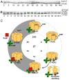
In vitro KaiC-P oscillation and biochemical model for KaiC-P oscillatory activity. (A) In vitro oscillation of KaiC-P for 4 days taken at 2-h time points. U-KaiC, unphosphorylated KaiC; KaiC-P, phosphorylated KaiC. (B) In vitro KaiC-P oscillation with resolved phosphoforms. In vitro reactions were incubated for 3 days with samples taken at 4-h time points. Four phosphoforms of KaiC exist over the course of a phosphorylation cycle. ST -KaiC, S431 and T432 KaiC double phosphoform; T-KaiC, T432 KaiC phosphoform; S-KaiC, S431 KaiC phosphoform; and U-KaiC, unphosphorylated KaiC. Immunoblot images provided by Yong-Ick Kim, UC-San Diego. (C) Model for ordered phosphorylation and monomer shuffling activity in KaiC. Initially at ZT 0, unphosphorylated KaiC (U-KaiC) associates with KaiA to induce phosphorylation of KaiC (T-KaiC) at T432 (white star) at ZT 4, which is subsequently followed by phosphorylation at S431 (black star) and possibly T426 (gray star) for a fully phosphorylated KaiC (ST-KaiC) by ZT 8. Fully phosphorylated KaiC initiates the dephosphorylation stage and is amenable to monomer shuffling between ZT 8 and 12 to synchronize multiple intracellular KaiC hexamers, leading to the lone S431 phosphoform (S-KaiC) by ZT 12 and association with KaiB. KaiC continues autodephosphorylation through ZT 16, yielding U-KaiC by ZT 20. Note that indicated KaiC phosphoforms at each ZT time represent the most prominent KaiC-P phosphoform relative to the population. P, phosphate.
Figure 3
Figure 3
Multiple roles for CikA in the S. elongatus circadian system. (A) Autophosphorylation of CikA at its histidine protein kinase (HPK) domain is positively influenced by the N-terminal GAF domain, but inhibited by the C-terminal pseudo-receiver (PsR) domain. (B) The PsR domain (circle) of CikA is proposed to interact with as-yet-to-be-identified proteins (triangles) at the pole of the cyanobacterial cell. Micrograph image of an S. elongatus cell harboring a ZsGreen-CikA fusion protein exhibits polar localization. Scale bar = 5μm. (C) Absence of the CikA-PsR domain results in the delocalization of this CikA variant to exist throughout the cytoplasm of the cell as shown in the cartoon and micrograph image, which visualizes the ZsGreen-CikAΔPsR fusion protein. Scale bar = 5μm. ZsGreen fusion constructs from Zhang et al., 1999; micrograph images courtesy of Julie Bordowitz, UC-San Diego. (D) CikA inhibits the ATPase activity of KaiC, such that in the absence of CikA, the ATPase activity rises above a maximum threshold to activate the SasA/RpaA two-component system of the circadian output pathway. RpaA is predicted to indirectly repress the localization of FtsZ protein that is involved in determining the midline of the cell to allow for division to proceed. (E) At ZT8, the nucleoid region of the wild-type (WT) and cikA null is diffuse. After being subjected to a 5-h dark pulse (DP5) beginning at ZT8, the cikA null does not display complete chromosome compaction like that of a wild-type cell.
Figure 4
Figure 4
Model of S. elongatus circadian output pathways. Internal timekeeping is mediated via regulation of KaiC ATPase and phosphorylation activities, where KaiA enhances KaiC autophosphorylation and KaiB averts the KaiA-induced stimulation. KaiC ATPase activity is repressed (indirectly) by CikA in a KaiA-independent pathway. Temporal information from the Kai complex is transduced through at least three protein-based pathways that regulate gene expression. KaiC stimulates SasA autophosphorylation; phosphorylated SasA subsequently transfers its phosphoryl group to RpaA, which leads to RpaA activation. This phosphotransfer event is predicted to result in changes in genome-wide gene expression patterns. LabA is involved in the negative regulation of RpaA function through indirect mechanisms. Residual rhythmic output in the absence of RpaA results from CikA-mediated gene expression pathways. KaiC activity also regulates chromosome compaction; a compact chromosome turns off expression of class 1 genes, while class 2 genes peak at times when compaction is at its highest. The exact mechanism by which KaiC influences chromosome topology is not yet known. Solid lines and dashed lines represent direct and indirect mechanisms, respectively. Arrows represent positive influence, while blunt lines represent inhibition. P, phosphate.

References

    1. Amdaoud M, Vallade M, Weiss-Schaber C, Mihalcescu I. Cyanobacterial clock, a stable phase oscillator with negligible intercellular coupling. Proc Natl Acad Sci USA. 2007;104:7051–6. - PMC - PubMed
    1. Arita K, Hashimoto H, Igari K, Akaboshi M, Kutsuna S, Sato M, Shimizu T. Structural and biochemical characterization of a cyanobacterium circadian clock-modifier protein. J Biol Chem. 2007;282:1128–35. - PubMed
    1. Aschoff J. Freerunning and entrained circadian rhythms. Handbook of behavioral neurobiology: Biological rhythms. 1981:81–93.
    1. Bell-Pedersen D, Cassone VM, Earnest DJ, Golden SS, Hardin PE, Thomas TL, Zoran MJ. Circadian rhythms from multiple oscillators: lessons from diverse organisms. Nat Rev Genet. 2005;6:544–56. - PMC - PubMed
    1. Chen TH, Chen TL, Hung LM, Huang TC. Circadian rhythm in amino acid uptake by Synechococcus RF-1. Plant Physiol. 1991;97:55–9. - PMC - PubMed

Publication types

Substances

LinkOut - more resources