

Actin cytoskeleton in myofibroblast differentiation: ultrastructure defining form and driving function
- PMID: 21925115
- PMCID: PMC3324184
- DOI: 10.1016/j.trsl.2011.05.004
Actin cytoskeleton in myofibroblast differentiation: ultrastructure defining form and driving function
Abstract
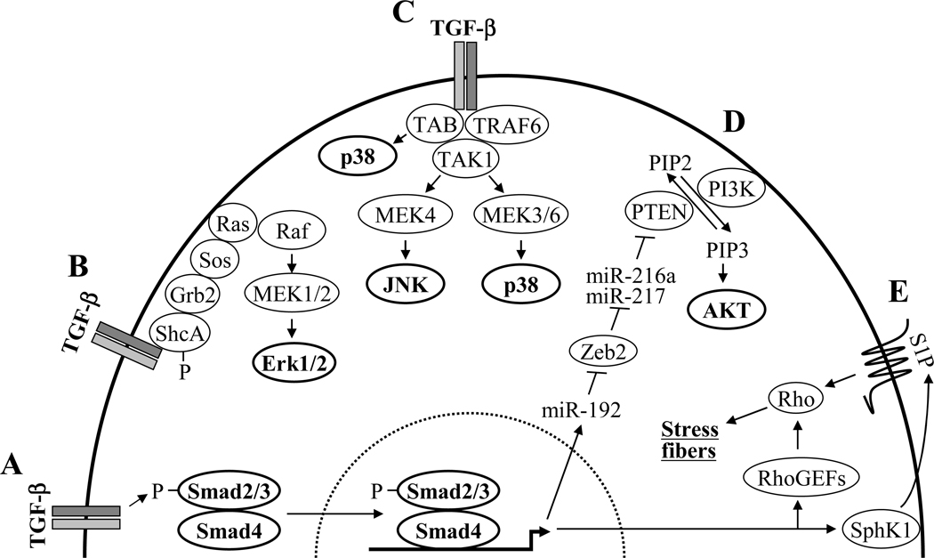
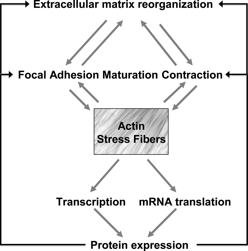
Myofibroblasts are modified fibroblasts characterized by the presence of a well-developed contractile apparatus and the formation of robust actin stress fibers. These mechanically active cells are thought to orchestrate extracellular matrix remodeling during normal wound healing in response to tissue injury; these cells are found also in aberrant tissue remodeling in fibrosing disorders. This review surveys the understanding of the role of actin stress fibers in myofibroblast biology. Actin stress fibers are discussed as a defining ultrastructural and morphologic feature and well-accepted observations demonstrating its participation in contraction, focal adhesion maturation, and extracellular matrix reorganization are presented. Finally, more recent observations are reviewed, demonstrating its role in transducing mechanical force into biochemical signals, transcriptional control of genes involved in locomotion, contraction, and matrix reorganization, as well as the localized regulation of messenger RNA (mRNA) translation. This breadth of functionality of the actin stress fiber serves to reinforce and amplify its mechanical function, via induced expression of proteins that themselves augment contraction, focal adhesion formation, and matrix remodeling. In composite, the functions of the actin cytoskeleton are most often aligned, allowing for the integration and amplification of signals promoting both myofibroblast differentiation and matrix remodeling during fibrogenesis.
Copyright © 2011 Mosby, Inc. All rights reserved.
Figures
References
-
- Gabbiani G, Ryan GB, Majne G. Presence of modified fibroblasts in granulation tissue and their possible role in wound contraction. Experientia. 1971;27:549–550. - PubMed
-
- Majno G, Gabbiani G, Hirschel BJ, Ryan GB, Statkov PR. Contraction of granulation tissue in vitro: similarity to smooth muscle. Science. 1971;173:548–550. - PubMed
-
- Ryan GB, Cliff WJ, Gabbiani G, Irle C, Montandon D, et al. Myofibroblasts in human granulation tissue. Hum Pathol. 1974;5:55–67. - PubMed
-
- Singer II. The fibronexus: a transmembrane association of fibronectin-containing fibers and bundles of 5 nm microfilaments in hamster and human fibroblasts. Cell. 1979;16:675–685. - PubMed
-
- Eyden B. The myofibroblast: an assessment of controversial issues and a definition useful in diagnosis and research. Ultrastruct Pathol. 2001;25:39–50. - PubMed
Publication types
MeSH terms
Substances
Grants and funding
LinkOut - more resources
Full Text Sources
