Review
. 2011 Sep 16;43(6):867-79.
doi: 10.1016/j.molcel.2011.08.024.

Prospects for riboswitch discovery and analysis

Affiliations
Review

Prospects for riboswitch discovery and analysis

Ronald R Breaker. Mol Cell. .

Abstract

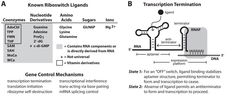
An expanding number of metabolite-binding riboswitch classes are being discovered in the noncoding portions of bacterial genomes. Findings over the last decade indicate that bacteria commonly use these RNA genetic elements as regulators of metabolic pathways and as mediators of changes in cell physiology. Some riboswitches are surprisingly complex, and they rival protein factors in their structural and functional sophistication. Each new riboswitch discovery expands our knowledge of the biochemical capabilities of RNA, and some give rise to new questions that require additional study to be addressed. Some of the greatest prospects for riboswitch research and some of the more interesting mysteries are discussed in this review.


Figures

Figure 1
Figure 1. Riboswitch ligands and mechanisms
(A) (Top) List of the riboswitch ligands with biochemical and or genetic validation. (Bottom) Demonstrated mechanisms for riboswitch-mediated gene control. (B) Schematic representation of the most common form of riboswitch-mediated gene regulation: transcription termination. Image depicts RNA polymerase (RNAP) in the act of transcribing the U-rich portion of an intrinsic transcription terminator stem in “State 1”, wherein ligand has been bound by the aptamer and transcription will terminate. Alternatively, in “State 2” (not shown), the absence of ligand allows nucleotides from the aptamer to form a competing anti-terminator stem that allows transcription to pass beyond the U-rich termination site. Other riboswitch mechanisms are described elsewhere (Breaker, 2011).
Figure 2
Figure 2. The collection of riboswitch classes that sense SAM and/or SAH
Depicted are the consensus sequence and structural models for four major families of riboswitches that respond to SAM. The SAM-I family is comprised of representatives classified as SAM-I (or S box), SAM-IV and SAM-I/IV riboswitches. Note that nucleotides shaded in turquoise are identical in these three consensus models, and these nucleotides are intimately involved informing the SAM-binding site (Montange and Batey, 2006). Similarly, the SAM-II family is comprised of SAM-II and SAM-V riboswitches that share nucleotides that form the ligand-binding pocket. The remaining two families are represented by SAM-III (or SMK box) and SAM-SAH riboswitch classes. A fifth riboswitch class that rejects SAM and selectively binds its metabolic derivative S-adenosylhomocysteine (SAH) (Wang et al., 2008) is not shown. P1 and related notations indicate base paired substructures.
Figure 3
Figure 3. Purine riboswitch variants and their ligand specificities
Depicted are the consensus sequence and secondary structure models for riboswitch aptamers that selectively respond to guanine, adenine or 2′-deoxyguanosine. Red nucleotides are present in greater than 90% of the guanine riboswitch representatives. Blue nucleotides in the adenine and 2′-deoxyguanosine aptamers differ from the guanine consensus. Other annotations are as described in the legend to Figure 2.
Figure 4
Figure 4. The most abundant orphan riboswitch classes
The consensus sequences and secondary structure models are depicted for the four most common classes of RNA elements believed to be riboswitch aptamers (Block et al., 2010; Meyer et al., 2011). (A) The yybP/ykoK motif RNAs are frequently associated with genes whose protein products mediate pH stress responses. (B) The ydaO/yuaA motif RNAs are frequently associated with genes involved in osmotic stress responses. (C) The pfl motif RNAs control genes centered on folate metabolism. (D) The ykkC/ykkD motif RNAs are associated with genes for transporters and for purine metabolism.
Figure 5
Figure 5. Architectures of riboswitches and their effects on ligand-mediated gene control
The expression platforms can be conventional systems as listed in Figure 1, or can be self-processing ribozymes. R designates the fraction of gene regulation, Y designates the fraction of aptamer bound by ligand, [L] designates ligand concentration, KD designates apparent dissociation constant. For all riboswitch architectures other than cooperative systems, values for Y are determined by using the equation for a simple riboswitch.
Figure 6
Figure 6. Allosteric control mechanism of a group I self-splicing ribozyme by a c-di-GMP riboswitch aptamer
Sequence and secondary structure model for the allosteric ribozyme from the bacterium C. difficile (Baker et al., 2010). A class II c-di-GMP riboswitch aptamer, formed by three conventional base-paired regions (P1 through P3) and one pseudoknot, resides only six nucleotides upstream of the initial structured portion (shaded P1) of a group I ribozyme. The 5′ splice site (red “5′ SS” between nucleotides 100 and 101) is a target for GTP attack (“GTP1”) when c-di-GMP is bound by the aptamer. Following this first step of splicing, the second step promotes attack by G101 of the 5′ exon at the 3′ splice site (red “3′ SS” between nucleotides 667 and 668) to yield spliced exons. In the absence of c-di-GMP binding, alternative base pairing (blue “anti-5′ SS stem) occurs between aptamer and ribozyme nucleotides. This anti-5′ SS stem displaces the ribozyme P1 stem, which precludes normal splicing. In contrast, an “alternative ribozyme P1 stem” (green) forms and promotes GTP attack (GTP2) after nucleotide 670. The products of normal splicing, promoted by c-di-GMP, are efficiently translated because they carry a typical ribosome binding site located the proper distance upstream of the unusual UUG start codon. The products of alternative splicing, which occurs when c-di-GMP is low in concentration, lack a ribosome binding site and therefore are not translated.

References

    1. Ames TD, Breaker RR. Bacterial riboswitch discovery and analysis. In: Meyer G, editor. The Chemical Biology of Nucleic Acids. John Wiley and Sons, Ltd; 2010. pp. 433–452.
    1. Ames TD, Breaker RR. Bacterial aptamers that selectively bind glutamine. RNA Biol. 2011;8:82–89. - PMC - PubMed
    1. Ataide SF, Wilson SN, Dand S, Rogers TE, Roy B, Banerjee R, Henkin TM, Ibba M. Mechanisms of resistance to an amino acid antibiotic that targets translation. ACS Chem Biol. 2007;2:819–827. - PMC - PubMed
    1. Batey RT, Gilbert SD, Montange RK. Structure of a natural guanine-responsive riboswitch complexed with the metabolite hypoxanthine. Nature. 2004;432:411–415. - PubMed
    1. Barrick JE, Corbino KA, Winkler WC, Nahvi A, Mandal M, Collins J, Lee M, Roth A, Sudarsan N, Jona I, Wickiser JK, Breaker RR. New RNA motifs suggest an expanded scope for riboswitches in bacterial gene control. Proc Natl Acad Sci USA. 2004;101:6421–6426. - PMC - PubMed

Publication types

LinkOut - more resources