. 2011 Sep 16;43(6):1040-6.
doi: 10.1016/j.molcel.2011.08.019.

The noncoding RNA Mistral activates Hoxa6 and Hoxa7 expression and stem cell differentiation by recruiting MLL1 to chromatin

Affiliations

The noncoding RNA Mistral activates Hoxa6 and Hoxa7 expression and stem cell differentiation by recruiting MLL1 to chromatin

Stéphane Bertani et al. Mol Cell. .

Retraction in

Abstract

The epigenetic activator Mixed lineage leukemia 1 (MLL1) is paramount for embryonic development and hematopoiesis. Here, we demonstrate that the long, noncoding RNA (lncRNA) Mistral (Mira) activates transcription of the homeotic genes Hoxa6 and Hoxa7 in mouse embryonic stem cells (mESC) by recruiting MLL1 to chromatin. The Mira gene is located in the spacer DNA region (SDR) separating Hoxa6 and Hoxa7, transcriptionally silent in mESCs, and activated by retinoic acid. Mira-mediated recruitment of MLL1 to the Mira gene triggers dynamic changes in chromosome conformation, culminating in activation of Hoxa6 and Hoxa7 transcription. Hoxa6 and Hoxa7 activate the expression of genes involved in germ layer specification during mESC differentiation in a cooperative and redundant fashion. Our results connect the lncRNA Mira with the recruitment of MLL1 to target genes and implicate lncRNAs in epigenetic activation of gene expression during vertebrate cell-fate determination.


Conflict of interest statement

The authors declare no competing financial interests.

Figures
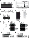
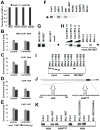
Figure 1
Figure 1. Identification and characterization of Mira
(A) Transcription of Hoxa genes, Glyceraldehyde-3-Phosphate Dehydrogenase (Gapdh), and Mira in undifferentiated (−RA), RA-treated (+RA), and control (differentiated in the absence of RA) mESCs detected by RvT-PCR. (B) RNA ChIP-on-chip assays detecting the interaction of Mll1 with chromatin-associated Mira in +RA mESCs. The relative abundance of RNAs, which associate with Mll1 in chromatin, is plotted over the corresponding template DNA of the Hoxa cluster (nucleotides 52135014–5214899) on chromosome 6. The position and transcriptional orientation (arrows) of Mira, Hoxa6, and Hoxa7 are indicated. (C) Structure of the Hoxa6/a7 cassette. Bars indicate the positions of probes in the Hoxa6/a7 cassette, which detected the Mira gene locus, an untranscribed region (IR), the promoter of Hoxa6 (PA6) and Hoxa7 (PA7), and the coding regions of Hoxa6 and Hoxa7 in ChIP assays. Arrows indicate the transcriptional start site of genes. (D) RNA ChIP assays detecting the association of Mll1 with Mira in native chromatin isolated from −RA, control, and +RA mESCs. (E) Primer extension assays detecting Mira transcript (arrowhead) in RNA isolated from +RA and −RA mESCs. (F) RvT-PCR assays detecting full-length Mira, an untranscribed region of the Hoxa6/a7-SDR (control) (Table S3), and Gapdh mRNA in control, −RA, and +RA mESCs. (G) Northern blot assays detecting Mira (arrowhead) in +RA and -RA mESCs. (D, E, G) The positions of size markers are indicated to the right. (H) ChIP assays detecting the presence of Mll1 and H3-K4(me3) at the Hoxa6/a7-SDR in chromatin isolated from −RA and +RA mESCs. Chromatin was precipitated with antibodies to Mll1, H3-K4(me3), or rabbit serum (mock). (I) ChIP assays detecting the association of the Mll1 complex, Menin, and LEDGF with the Mira gene in −RA, +RA mESCs, and +RA mESCs lacking Menin or Mll1 through RNAi. Chromatin was precipitated with antibodies to the indicated antigens and rabbit serum (mock). (J) RNase-ChIP assays detecting the association of Mll1 with the Hoxa6/a7 cassette. Chromatin was isolated from +RA mESCs and treated with BSA and RNase inhibitor (mock), RNase A, or RNase H (K) ChIP assays as in (I) except that RNase H treated chromatin was used. See also Figure S1, S2, and S3.
Figure 2
Figure 2. Mll1 interacts with Mira
(A) Nuclear run-on assays detecting Mira, Hoxa6, Hoxa7, and Gapdh transcription in nuclei of +RA mESCs, which had been incubated with tetracycline (mock) or α-amanitin. (B) RNA ChIP assays detecting the association of Mll1 with Mira in native chromatin of +RA mESCs, which had been treated with tetracycline (mock) or α-amanitin. Native chromatin was immunoprecipitated with antibodies to Mll1, H3K4 (me3), and rabbit serum (mock). (C–E) ChIP assays detecting the association of Mll1 with the Mira gene locus (C) and the promoter of Hoxa6 (D) and Hoxa7 (E) in +RA cells described in (B). (A–E) Error bars represent the standard error of the mean (SEM). (F) In vitro protein-RNA binding assays detecting the interaction of Mira with endogenous Mll1 (eMll1), recombinant MLL1C180, Mll1ΔSET, which lacks the SET-domain, WDR5, Ash2L, RpBP5, LEDGF, and Menin. (G) In vitro binding assays detecting the interaction of Mira with recombinant Mll1SET, Mll1 ΔSET, Mll1C180, and Ash1SET (mock). (H) In vitro binding assays detecting the interaction of sense (+) and anti-sense (−) Mira with Mll1 ΔSET and Ash1SET (mock). (I) In vitro binding assays detecting the interacting of truncated Mira transcripts with Mll1SET or Ash1SET (mock). (J) Schematic representation of (top) Mira and (bottom) wild type and mutant ABM. (K) In vitro binding assays detecting the interaction of Mll1SET, Mll1SET(G3836S) and Mll1(R3880A) with wild type and mutant ABM. (F, H, I, K) Input represents 15% of the radiolabeled RNA present in in vitro binding assays. See also Figure S2 and S3.
Figure 3
Figure 3. Mira mediated recruitment of Mll1 promotes transcription of Hoxa6 and Hoxa7
(A) Transcription of Mira, Hoxa6, Hoxa7, and Gapdh in +RA mESCs treated with control siRNA (mock-siRNA) or siRNA targeting Mira (Mira-siRNA1). (B) Transcription of Mira, Gapdh, and Hoxa genes in mock-siRNA and Mira-siRNA1 mESCs. (C) ChIP assays monitoring the association of Mll1 and a control (rabbit serum) with the Hoxa6/a7 cassette and Hoxa genes in mock-siRNA and Mira-siRNA1 mESCs. Target DNA regions are described in Figure 2A. (D) ChIP assays as described in (C) detecting the association of H3K4(me3) with the Hoxa6/a7 cassette. (E) Schematic representation of the Hoxa6/a7 cassette. The positions of PCR primer pairs used for 3C assays are indicated. (F) 3C assays detecting the association of the Mira gene locus with Hoxa6 and Hoxa7 in (top panel) −RA and +RA mESCs, and (bottom panel) +RA mESCs, which contain (+) or lack Mira through RNAi (−). (G) Binding of Mll1SET, Mll1SET(G3836S), and Mll1SET(R3880A) to RNA/DNA complexes containing ssDNA (black) and truncated Mira (dark grey) containing a wild type or mutant ABM. See also Figure S3. (H) In vitro protein nucleic acid binding assays as described in (F) except that the RNA/DNA hybrids were preincubated with RNase-A or -H.
Figure 4
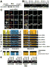
Figure 4. Mira controls the expression of germ layer marker genes
(A) Schematic representation of the Mira gene locus. Black boxes indicate the positions of Mira-siRNA1 and Mira-siRNA2. (B) Transcription of Mira, Hoxa6, Hoxa7, and Gapdh in +RA mock-siRNA, Mira-siRNA1 and Mira-siRNA2 mESCs. (C) FISH coupled to immunofluorescence (IF) assays detecting Mira and Nestin together with T Sox17, Hox-A6, or Hox-A7 in +RA mock-siRNA and +RA Mira-siRNA1 cells. (D–F) Transcription of germ layer marker genes in (D) mock-siRNA, (E) Mira-siRNA1, and (F) Mira-siRNA2 +RA mESCs. Target genes for Mira are highlighted in yellow (ectoderm), blue (endoderm), and orange (mesoderm). (G–I) Marker gene transcription in +RA mESCs, which had been transfected with (G) Hoxa6-siRNA, (H) Hoxa7-siRNA, and (I) Hoxa6 + Hoxa7 siRNA. (J) Results of the RvT-PCR assays described in (D-I). Dark boxes indicate target genes for Mira. The numbers 6 and 7 indicate the target genes for Hoxa6 and Hoxa7, respectively. The symbols (67R) and (67C) indicate genes activated by Hoxa6 and Hoxa7 in a redundant fashion and cooperative, respectively, fashion. For gene nomenclature see Table S1. (K) RvT-PCR assays detecting the transcription of Mira, Hoxa6, Hoxa7, and Gapdh in mock-siRNA and +RA mESCs, which lack Hoxa6 (Hoxa6-siRNA), Hoxa7 (Hoxa7-siRNA), or Hoxa6 and Hoxa7 (Hoxa6 + Hoxa7 siRNA) through RNAi. See also Fig. S4.

References

    1. Bertani S, Kan A, Sauer F. Chromatin Immunoprecipitation from human embryonic stem cells. J Vis Exp. 2008 doi: 10.3791/780. - DOI - PMC - PubMed
    1. Boyer LA, Plath K, Zeitlinger J, Brambrink T, Medeiro LA, Lee TI, Levine SS, Wernig M, Tajonar A, Ray MK, et al. Polycomb complexes repress developmental regulators in murine embryonic stem cells. Nature. 2006;441:349–353. - PubMed
    1. Bracken AP, Dietrich N, Pasini D, Hansen KH, Helin K. Genome-wide mapping of Polycomb target genes unravels their roles in cell fate transitions. Genes Dev. 2006;20:1123–1136. - PMC - PubMed
    1. Carroll SB. Homeotic genes and the evolution of arthropods and chordates. Nature. 1995;376:479–485. - PubMed
    1. Cosgrove MS, Patel A. Mixed lineage leukemia: a structure-function perspective of the MLL1 protein. FEBS J. 2010;277:1832–1842. - PMC - PubMed

Publication types

MeSH terms

Associated data