. 2011 Dec;141(6):2109-18.
doi: 10.1053/j.gastro.2011.09.015. Epub 2011 Sep 16.

Gut-tropic T cells that express integrin α4β7 and CCR9 are required for induction of oral immune tolerance in mice

Affiliations

Gut-tropic T cells that express integrin α4β7 and CCR9 are required for induction of oral immune tolerance in mice

Barbara Cassani et al. Gastroenterology. 2011 Dec.

Abstract

Background & aims: Induction of oral immune tolerance (OT) blocks proinflammatory responses to orally administered antigens and might be used to treat autoimmune conditions. We investigated whether gut-tropic T cells that express the integrin α4β7 and the chemokine receptor CCR9 are required for OT.

Methods: Skin delayed-type hypersensitivity and experimental autoimmune encephalomyelitis were used to monitor OT in mice. To assess the role of receptors that mediate localization of lymphocytes to the gut (gut-homing receptors) in induction of OT, we studied CCR9(-/-) and β7(-/-) mice and also blocked the α4β7 ligand MAdCAM-1 in wild-type mice. We used DEREG and Scurfy mice to assess the role of Foxp3(+) regulatory T cells (Treg) and IL-10(-/-) and IL-10Rβ(-/-) mice to examine the role of interleukin (IL)-10 in induction of OT.

Results: OT could not be induced in CCR9(-/-) or β7(-/-) mice, or when MAdCAM-1 was blocked in wild-type mice, indicating that gut-homing receptors are required for oral tolerization. Consistent with the role of all-trans retinoic acid in inducing gut-homing T cells, OT could not be induced in mice depleted of vitamin A. OT was rescued in CCR9(-/-) mice following adoptive transfer of wild-type T cells, but not CCR9(-/-) or β7(-/-) T cells. Gut-homing T cells are therefore necessary and sufficient to induce OT. Wild-type Treg and IL-10 were required to restore OT to CCR9(-/-) mice, indicating that homing and functional differentiation of IL-10-producing Treg in the gut is required for OT. Conversely, transfer of CCR9(-/-) or β7(-/-) T cells to wild-type mice partially inhibited OT.

Conclusions: Expression of CCR9 and α4β7 on T cells and their subsequent localization to the gut is required for induction of OT in mice. Therapies designed to block gut-homing receptors might, under some conditions, interfere with normal tolerogenic mechanisms in the intestinal mucosa.


Figures

Figure 1
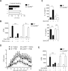
Figure 1. CCR9 is required for OT induction
(A–C) Wild type (wt) and CCR9−/− mice were supplemented daily with oral PBS or 2.5 mg OVA for 5 days, and then they were immunized s.c. with OVA plus CFA. After 7 days the mice were challenged with OVA in the left footpad and PBS in the right footpad. (A) DTH responses were measured after 24 hours (n=7–9 mice/group). (B, C) Total splenocytes were stimulated with 1 mg/ml OVA. (B) Cell proliferation was assessed after 72 hours. (C) Cytokines were measured in the culture supernatant after 24–48 hours. (D, E) Wt and CCR9−/− mice were supplemented daily with oral PBS or 0.25 mg MOG35–55 for 5 days, then immunized s.c. with MOG35–55 plus CFA. (D) EAE induction and progression was scored from days 11 to 25 (n=8–10 mice/group). (E) At day 16, the mice were sacrificed and total splenocytes were stimulated ex vivo with 20 μg/ml MOG35–55. T cell proliferation was measured after 72 hours (n=4 mice/group). Mean ± SEM, *p<0.05, **p<0.01, ***p<0.001, n.s.: not significant.
Figure 2
Figure 2. Blocking MAdCAM-1 abrogates OT in wt mice
Wt mice were treated i.p. with anti-MAdCAM-1 or control rIgG. OT and DTH were induced as described in Fig. 1. (A) Flow cytometry analysis of one representative mouse/group. (B) Frequencies of α4β7- and CCR9-expressing cells in the spleen. Cells were gated on CD44HiCD62LLow effector/memory CD4+ T cells (C) DTH responses were measured after 24 hours (n=2–3 mice/group). (D, E) Total splenocytes were stimulated with the indicated OVA concentrations. (D) Proliferation was measured after 72 hours (n=2–3 mice/group). (E) Cytokines were measured in the culture supernatant after 24–48 hours. Mean ± SEM, *P<0.05, *P<0.01,***P<0.001, n.s.: not significant.
Figure 3
Figure 3. OT is abolished in vitamin A depleted (VAD) mice
Mice were maintained on a control or VAD diet. OT and DTH were induced as described in Fig. 1. (A) DTH responses (footpad swelling) were measured after 24 hours, and were expressed as the difference between the left and right footpad (L-R) (n=4 mice/group). (B, C) Total splenocytes were stimulated with 1 mg/ml OVA. (B) T cell proliferation was measured after 72 hours. (C) Cytokines were measured in the culture supernatant after 24–48 hours. Mean ± SEM, *p<0.05, **p<0.01, ***p<0.001, n.s.: not significant.
Figure 4
Figure 4. Adoptive transfer of wt T cells rescues OT in CCR9−/− mice
T cells from nontolerized wt or CCR9−/− mice were adoptively transferred into CCR9−/− recipients. OT and DTH were induced as described in Fig. 1. CCR9−/− mice receiving CD4+ T cells from DEREG mice were treated with DT every day. (A, E) DTH responses were measured after 24 hours (n=2–5 mice/group). (B–D, F) Total splenocytes were stimulated with the indicated OVA concentrations. (B, D, F) Proliferation was assessed after 72 hours (n=4–5 mice/group). (C) IL-10 was measured in the culture supernatant after 48–60 hours. Mean ± SEM, *p<0.05, ***p<0.001
Figure 5
Figure 5. IL-10 signals in gut-tropic TREG are critical for OT
T cells from IL-10−/− or IL-10Rβ−/− mice were adoptively transferred into CCR9−/− mice. OT and DTH were induced as described in Fig. 1. (A) Total splenocytes were stimulated with the indicated OVA concentrations and cell proliferation was measured after 72 h. (B, C) Cytokines were measured in the culture supernatant after 48–60 hours. n=2 mice/group, Mean ± range
Figure 6
Figure 6. Transfer of CCR9−/− or β7−/− T cells impairs OT in wt mice
T cells from CCR9−/− or β7−/− mice were adoptively transferred into wt mice. OT and DTH were induced as described in Fig. 1. (A) DTH responses were measured after 24h (n= 2 mice/group). Groups were compared versus wild type mice receiving oral PBS. (B) Total splenocytes were stimulated with the indicated OVA concentrations and cell proliferation was measured after 72h (n= 2 mice/group). Mean ± SEM, *p<0.05 (ANOVA)
Figure 7
Figure 7. Gut-homing-dependent model for OT
(A) The prevailing view indicates that OT relies on the induction of antigen-specific Foxp3+ regulatory T cells (TREG) in the mesenteric lymph nodes (MLN), which then reach the blood and suppress pro-inflammatory responses in different tissues. (B) In our new model we propose that TREG require a second activation step in the intestinal lamina propria (LP) to complete their differentiation into fully tolerogenic IL-10-producing TREG, which can then suppress pro-inflammatory responses. While the first step in MLN involves naïve T cells and therefore does not require gut-homing receptors, the second step in the gut LP depends on the induction of retinoic acid (RA)-dependent gut-homing receptors α4β7 and CCR9 on activated TREG and their respective ligands MAdCAM-1 and CCL25 on LP endothelial cells. In the LP TREG are re-stimulated and proliferate in response to oral antigen and they are also exposed to IL-10, which is required to confer TREG with IL-10-producing capacity, a critical property for OT generation. After that, IL-10+ TREG return to the blood and exert their tolerogenic activity in different organs.

References

    1. Chen Y, Inobe J, Marks R, Gonnella P, Kuchroo VK, Weiner HL. Peripheral deletion of antigen-reactive T cells in oral tolerance. Nature. 1995;376:177–80. - PubMed
    1. Dubois B, Joubert G, Gomez de Aguero M, Gouanvic M, Goubier A, Kaiserlian D. Sequential role of plasmacytoid dendritic cells and regulatory T cells in oral tolerance. Gastroenterology. 2009;137:1019–28. - PubMed
    1. Zhang X, Izikson L, Liu L, Weiner HL. Activation of CD25(+)CD4(+) regulatory T cells by oral antigen administration. J Immunol. 2001;167:4245–53. - PubMed
    1. Spahn TW, Weiner HL, Rennert PD, Lugering N, Fontana A, Domschke W, Kucharzik T. Mesenteric lymph nodes are critical for the induction of high-dose oral tolerance in the absence of Peyer's patches. Eur J Immunol. 2002;32:1109–13. - PubMed
    1. Worbs T, Bode U, Yan S, Hoffmann MW, Hintzen G, Bernhardt G, Forster R, Pabst O. Oral tolerance originates in the intestinal immune system and relies on antigen carriage by dendritic cells. J Exp Med. 2006;203:519–27. - PMC - PubMed

Publication types

MeSH terms