. 2011 Oct 28;413(3):712-25.
doi: 10.1016/j.jmb.2011.08.059. Epub 2011 Sep 8.

Structural characterization of the interactions between palladin and α-actinin

Affiliations

Structural characterization of the interactions between palladin and α-actinin

Moriah R Beck et al. J Mol Biol. .

Abstract

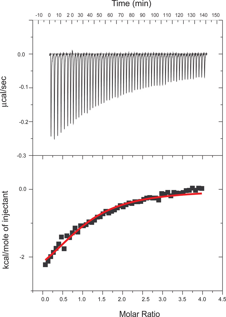
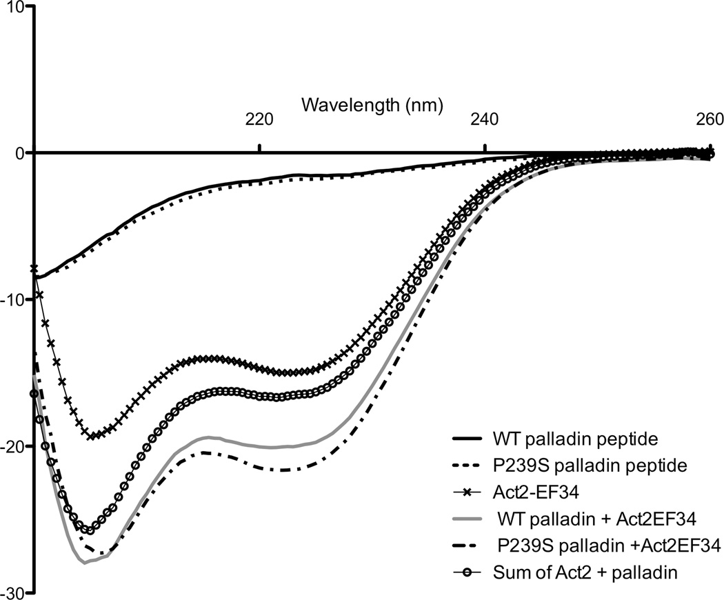
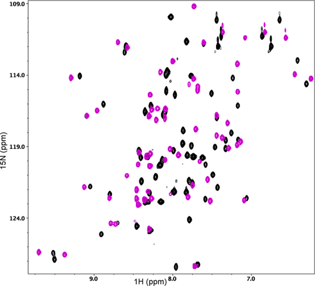
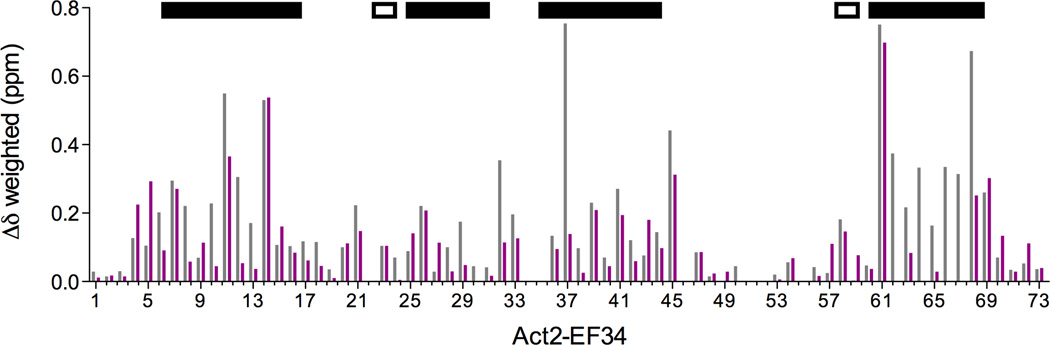
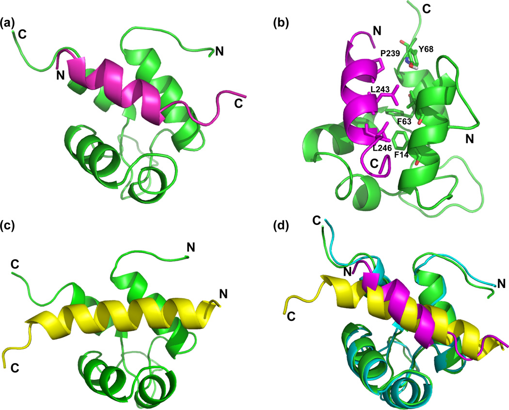
The interaction between α-actinin and palladin, two actin-cross-linking proteins, is essential for proper bidirectional targeting of these proteins. As a first step toward understanding the role of this complex in organizing cytoskeletal actin, we have characterized binding interactions between the EF-hand domain of α-actinin (Act-EF34) and peptides derived from palladin and generated an NMR-derived structural model for the Act-EF34/palladin peptide complex. The critical binding site residues are similar to an α-actinin binding motif previously suggested for the complex between Act-EF34 and titin Z-repeats. The structure-based model of the Act-EF34/palladin peptide complex expands our understanding of binding specificity between the scaffold protein α-actinin and various ligands, which appears to require an α-helical motif containing four hydrophobic residues, common to many α-actinin ligands. We also provide evidence that the Family X mutation in palladin, associated with a highly penetrant form of pancreatic cancer, does not interfere with α-actinin binding.


Figures

Figure 1
Figure 1
ITC binding isotherm obtained for the interaction of palladin (WT) with Act-EF34. Fifty-five 5 µL aliquots of palladin (1 mM) were injected into the calorimeter cell containing Act-EF34 (600 µM) at 27 °C.
Figure 2
Figure 2
Gain of signal for the Act-EF34/palladin complex recorded by far-UV CD spectroscopy. The concentration of each component was 20 µM, and the path length was 2 mm. The spectra of the isolated components, WT palladin (—), FX palladin (- -), Act2-EF34 (✕), WT complex (—), and FX complex (–·-) are shown. The sum of the spectra for the isolated Act-EF34 and palladin peptide is also reported for comparison (○).
Figure 3
Figure 3
(a) 2D 1H-15N HSQC spectral overlay of 15N-labeled Act-EF34 in the absence (black) and presence of bound WT palladin peptide (magenta). (b) Chemical shift perturbation of Act-EF34 amide resonances upon complex formation with either titin Zr7 (gray) or palladin peptide (magenta). The α-helical regions or β-strands of Act-EF34 are designated by filled or open bars, respectively, above the plot. Also indicated (dotted line) is the chemical shift cut-off value used for the definition of AIRs as input for the HADDOCK calculations (0.17 ppm).
Figure 3
Figure 3
(a) 2D 1H-15N HSQC spectral overlay of 15N-labeled Act-EF34 in the absence (black) and presence of bound WT palladin peptide (magenta). (b) Chemical shift perturbation of Act-EF34 amide resonances upon complex formation with either titin Zr7 (gray) or palladin peptide (magenta). The α-helical regions or β-strands of Act-EF34 are designated by filled or open bars, respectively, above the plot. Also indicated (dotted line) is the chemical shift cut-off value used for the definition of AIRs as input for the HADDOCK calculations (0.17 ppm).
Figure 4
Figure 4
Ligand binding alters the backbone flexibility of Act2-EF34 as monitored by NMR relaxation analyses. Relaxation parameters, (a) {1H}-15N NOE values and (b) the generalized order parameter (S2) for Act-EF34 in both its palladin-bound (filled circles) and free (open circles) forms, plotted against α-actinin residue number.
Figure 5
Figure 5
Comparison of titin-bound versus palladin-bound Act-EF34 revealed by residual dipolar coupling NMR (RDC) data. (a) Correlation plot of experimentally measured RDCs for Act-EF34 bound to palladin peptide versus back-calculated RDCs based on the NMR structure of Act-EF34 bound to titin Zr7 (PDB code: 1H8B) using REDCAT. (b) The absolute difference between the observed and expected dipolar couplings (ΔRDC) is plotted as a function of amino acid residue for Act-EF34.
Figure 6
Figure 6
Comparison of the Act-EF34 complex with palladin and titin peptides. (a) Ribbon diagram showing a representative palladin complex structural model. Act-EF34 is shown in green and palladin in magenta, with termini labeled to highlight orientation. (b) Critical residues used in docking are highlighted. The backbone atoms associated with the hydrophobic Act-EF34 Phe 14, Phe 63, and Tyr 68 residues that were utilized as restraints in docking. Palladin residues, Leu 243 and 246, are hydrophobic residues in the first and fourth position of the ‘1-4-5-8’ motif. These residues were mutated, found to abolish binding to Act-EF34, and then used as restraints for docking. (c) For comparison, the ribbon diagram of the complex between Act-EF34 (green) and titin’s seventh Z-repeat in yellow is shown (Act-EF34—Zr7; PDB ID: 1H8B). (d) Structural alignment of palladin-bound (green with magenta peptide) and titin-bound (cyan with yellow) Act-EF34.

References

    1. Revenu C, Athman R, Rovine S, Louvard D. The co-workers of actin filaments: from cell structures to signals. Nat Rev Mol Cell Biol. 2004;5:635–646. - PubMed
    1. Otey CA, Carpen O. alpha-Actinin revisited: a fresh look at an old player. Cell Motil Cytoskeleton. 2004;58:104–111. - PubMed
    1. Parast MM, Otey CA. Characterization of palladin, a novel protein localized to stress fibers and cell adhesions. J Cell Biol. 2000;150:643–656. - PMC - PubMed
    1. Mykkanen OM, Gronholm M, Ronty M, Lalowski M, Salmikangas P, Suila H, Carpen O. Characterization of human palladin, a microfilament-associated protein. Mol Biol Cell. 2001;12:3060–3073. - PMC - PubMed
    1. Boukhelifa M, Parast MM, Bear JE, Gertler FB, Otey CA. Palladin is a novel binding partner for Ena/VASP family members. Cell Motil Cytoskeleton. 2004;58:17–29. - PubMed

Publication types