. 2011 Nov 18;286(46):40276-86.
doi: 10.1074/jbc.M111.283671. Epub 2011 Sep 16.

Charged multivesicular body protein 2B (CHMP2B) of the endosomal sorting complex required for transport-III (ESCRT-III) polymerizes into helical structures deforming the plasma membrane

Affiliations

Charged multivesicular body protein 2B (CHMP2B) of the endosomal sorting complex required for transport-III (ESCRT-III) polymerizes into helical structures deforming the plasma membrane

Gilles Bodon et al. J Biol Chem. .

Abstract

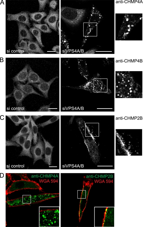
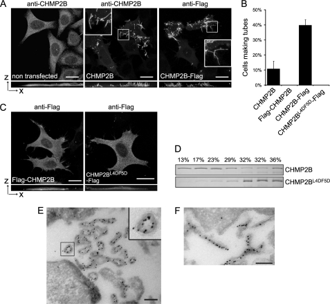
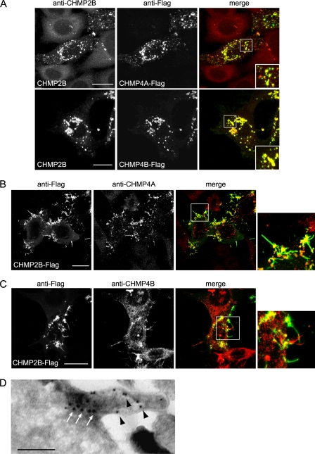
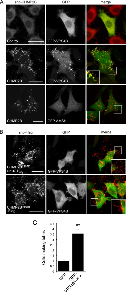
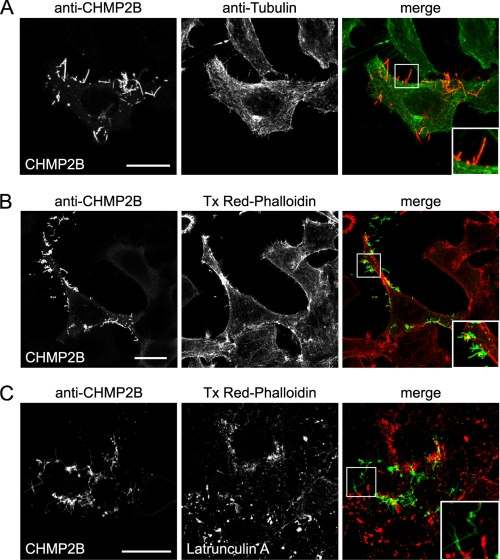
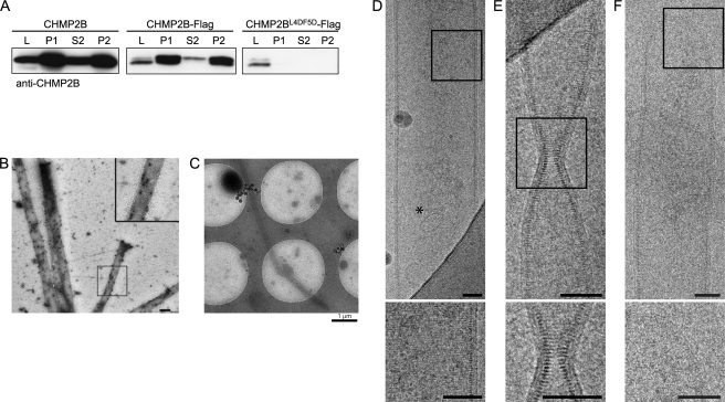
The endosomal sorting complexes required for transport (ESCRT-0-III) allow membrane budding and fission away from the cytosol. This machinery is used during multivesicular endosome biogenesis, cytokinesis, and budding of some enveloped viruses. Membrane fission is catalyzed by ESCRT-III complexes made of polymers of charged multivesicular body proteins (CHMPs) and by the AAA-type ATPase VPS4. How and which of the ESCRT-III subunits sustain membrane fission from the cytoplasmic surface remain uncertain. In vitro, CHMP2 and CHMP3 recombinant proteins polymerize into tubular helical structures, which were hypothesized to drive vesicle fission. However, this model awaits the demonstration that such structures exist and can deform membranes in cellulo. Here, we show that depletion of VPS4 induces specific accumulation of endogenous CHMP2B at the plasma membrane. Unlike other CHMPs, overexpressed full-length CHMP2B polymerizes into long, rigid tubes that protrude out of the cell. CHMP4s relocalize at the base of the tubes, the formation of which depends on VPS4. Cryo-EM of the CHMP2B membrane tubes demonstrates that CHMP2B polymerizes into a tightly packed helical lattice, in close association with the inner leaflet of the membrane tube. This association is tight enough to deform the lipid bilayer in cases where the tubular CHMP2B helix varies in diameter or is closed by domes. Thus, our observation that CHMP2B polymerization scaffolds membranes in vivo represents a first step toward demonstrating its structural role during outward membrane deformation.


Figures

FIGURE 1.
FIGURE 1.
VPS4A and -B down-regulation induces plasma membrane accumulation of endogenous CHMP2B. HeLa cells were depleted of VPS4A and VPS4B using a mixture of siRNA against both proteins (right panels). Control cells were transfected with an irrelevant siRNA (left panels). The confocal section of control cells or of cells depleted of VPS4 was immunostained with antibodies against CHMP4A (A), CHMP4B (B), or CHMP2B (C). Enlargements of the boxed regions are shown on the right. D, confocal sections of VPS4-depleted cells treated with Alexa Fluor 594-WGA to delineate membranes. Cells were immunostained using anti-CHMP4A (left panel) or anti-CHMP2B (right panel). Scale bars, 20 μm.
FIGURE 2.
FIGURE 2.
Overexpression of CHMP2B or CHMP2B-FLAG induces formation of long cell surface protrusions in which they concentrate. HeLa cells were transfected with the indicated plasmids and immunostained 36 h later. Except otherwise stated, photographs presented in all figures are maximal intensity projections of confocal image stacks. In A and C, bottom panels represent projections in the x-z plane. Bars, 20 μm. A, CHMP2B immunostaining of cells transfected with the empty vector reveals a homogeneous cytoplasmic localization of the protein. Overexpression of CHMP2B and CHMP2B-FLAG induces the formation of cell surface protrusions in which CHMP2B concentrates. Note the absence of cytoplasmic staining in tube forming cells. B, percentage of transfected cells displaying tubes as revealed with anti-CHMP2B antibodies. For each condition, 200 CHMP2B-expressing cells were counted. Data from three independent experiments are plotted. Standard error bars are shown. C, FLAG-CHMP2B and CHMP2BL4D/F5D-FLAG do not induce surface protrusions. D, flotation experiments demonstrate that mutations in Leu-4–Phe-5 impair the capacity of CHMP2B to associate with membranes in vitro. Purified recombinant full-length CHMP2B and CHMP2BL4D/F5D were mixed with liposomes. Liposomes were floated on a sucrose gradient, fractions were run on SDS-PAGE, and proteins were revealed by Coomassie staining. E and F, FLAG immunogold labeling of cells overexpressing CHMP2B-FLAG. Cross-sections of tubes reveal the presence of the C-terminal FLAG lining the lumen of a hollow, electron-dense structure closely associated with the membrane of tubes; longitudinal sections reveal the presence of CHMP2B along the entire length of the tubes. Scale bars, 200 nm.
FIGURE 3.
FIGURE 3.
Relationship between CHMP4- and CHMP2B-induced protrusions. HeLa cells were transfected with the indicated plasmids and immunostained 36 h later. A, CHMP4A-FLAG or CHMP4B-FLAG, co-expressed with similar amounts of CHMP2B, block the capacity of CHMP2B to form protrusions. Note that the relocalization of CHMP2B inside CHMP4A or CHMP4B patches. In cells expressing CHMP2B-FLAG only, immunostaining with anti-CHMP4A (B) or anti-CHMP4B (C) antibodies reveals the relocalization of endogenous CHMP4A and CHMP4B at the base of CHMP2B tubes. The right panels show enlargement of the boxed regions in the merged panels. In all cases, scale bars are 20 μm. D, double immunogold labeling of a tube emanating from a cell overexpressing CHMP2B-FLAG. Anti-FLAG staining (10-nm beads, black arrowheads) reveals the presence of CHMP2B along the entire length of the tube; anti CHMP4A (15-nm beads, white arrows) reveals CHMP4A labeling restricted at the base of the tube. Scale bars, 200 nm.
FIGURE 4.
FIGURE 4.
Relationship between VPS4 and CHMP2B protrusions. HeLa cells were co-transfected with the indicated plasmids and observed 36 h later. A, in cells transfected with the control plasmid together with GFP-VPS4, VPS4 is distributed homogeneously in the cytoplasm. In cells expressing both GFP-VPS4B and CHMP2B, VPS4 is mainly present in CHMP2B tubes. In contrast, AMSH is not recruited in CHMP2B protrusions made by cells expressing both GFP-AMSH and CHMP2B. B, CHMP2BL207D/L210D-FLAG, mutated in the MIM domain, accumulates in plasma membrane patches but does not induce protrusions. CHMP2Bintron5, which lacks the C-terminal α6 helix containing the MIM domain, forms everting tubes. GFP-VPS4 is recruited neither in CHMP2BL207D/L210D-FLAG patches nor in CHMP2Bintron5-FLAG containing tubes. C, the catalytically inactive form of VPS4B (GFP-VPS4BE235Q) increases the proportion of cells displaying CHMP2B protrusions: HeLa cells were transfected with CHMP2B together with GFP-VPS4BE235Q and observed 24 h later. For each condition, 200 cells from three independent experiments were counted. Standard error bars are shown. Mann-Whitney statistical analysis was used (p < 0.0011). Scale bars, 20 μm. **, p < 0.0011.
FIGURE 5.
FIGURE 5.
HeLa cells CHMP2B protrusions do not contain actin or tubulin. Immunostaining with anti-β-tubulin antibody (A) and staining with Texas Red (Tx Red)-phalloidin (B) of HeLa cells overexpressing CHMP2B demonstrate the absence of tubulin and F-actin in CHMP2B tubes. C, latrunculin A treatment (0.1 μg/ml; 60 min) induces depolymerization of actin but does not collapse CHMP2B tubes.
FIGURE 6.
FIGURE 6.
Characterization of tubes pelleted from culture media of CHMP2B-expressing cells. A, Western blotting analysis using anti-CHMP2B of culture media of CHMP2B, CHMP2B-FLAG, and CHMP2BL4D/F5D-FLAG expressing cells. L, cell lysates; P1, 30,000 × g pellet of culture media. P2 and S2, P1 pellets solubilized in 1% Triton X-100 were centrifuged at 20,000 × g: most of the CHMP2B-containing material present in culture media is resistant to detergent extraction. No CHMP2B pelletable material could be recovered from media of CHMP2BL4D/F5D-FLAG-expressing cells. B, P1 pellets of culture medium of CHMP2B-FLAG-expressing cells contain tubes made up of CHMP2B. P1 pellets were fixed, permeabilized, and immunolabeled with anti-CHMP2B antibodies revealed by protein A gold (10 nm). C–F, cryo-EM analysis of P1 pellets prepared from culture medium from CHMP2B-FLAG-expressing cells reveals the structure of CHMP2B tubes. C, low magnification shows tubes with a length reaching at least 8 μm. D, striations perpendicular to the longitudinal axis can be seen across the width of the tube. An asterisk indicates a 50-nm vesicle inside of the tube. E, constriction of a tube with no change in the helix pitch. F, dome closing one end of a tube. The inner leaflet of the membrane is closely associated with the CHMP2B protein lattice. Scale bars, 50 nm.

References

    1. Babst M., Katzmann D. J., Snyder W. B., Wendland B., Emr S. D. (2002) Dev. Cell 3, 283–289 - PubMed
    1. Babst M., Katzmann D. J., Estepa-Sabal E. J., Meerloo T., Emr S. D. (2002) Dev. Cell 3, 271–282 - PubMed
    1. Katzmann D. J., Babst M., Emr S. D. (2001) Cell 106, 145–155 - PubMed
    1. Hurley J. H. (2010) Crit. Rev. Biochem. Mol. Biol. 45, 463–487 - PMC - PubMed
    1. Shestakova A., Hanono A., Drosner S., Curtiss M., Davies B. A., Katzmann D. J., Babst M. (2010) Mol. Biol. Cell 21, 1059–1071 - PMC - PubMed

Publication types

MeSH terms

Substances