. 2011 Nov;193(22):6276-87.
doi: 10.1128/JB.05899-11. Epub 2011 Sep 16.

CodY-mediated regulation of guanosine uptake in Bacillus subtilis

Affiliations

CodY-mediated regulation of guanosine uptake in Bacillus subtilis

Boris R Belitsky et al. J Bacteriol. 2011 Nov.

Abstract

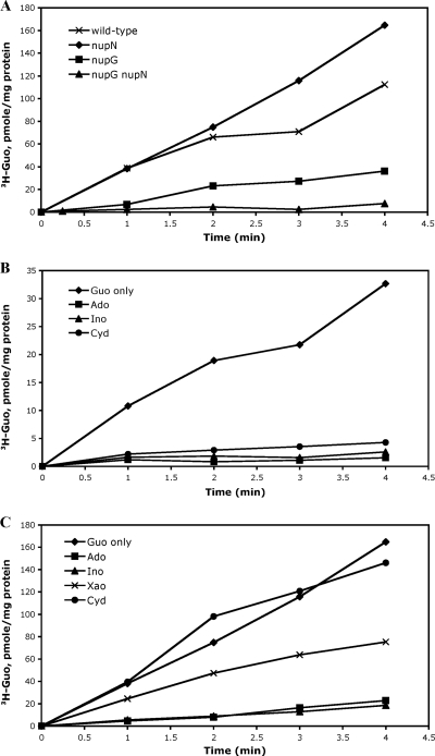
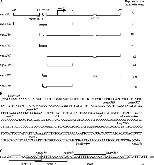
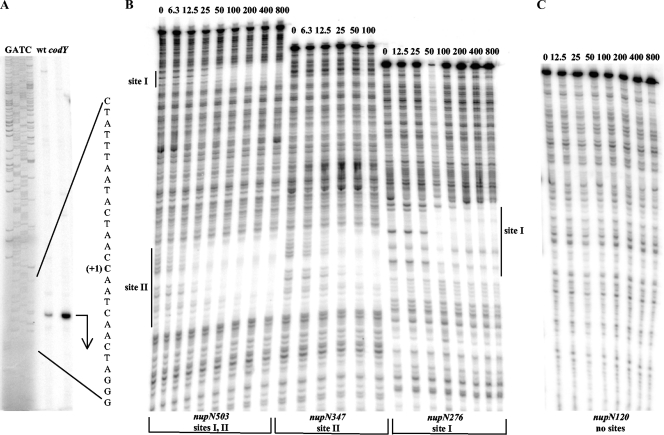
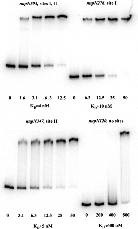
CodY is a global transcriptional regulator known to control expression of more than 100 genes and operons in Bacillus subtilis. Some of the most strongly repressed targets of CodY, the nupNOPQ (formerly, yufNOPQ) genes, were found to encode a guanosine transporter. Using DNase I footprinting experiments, we identified two high-affinity CodY-binding sites in the regulatory region of the nupN gene. The two sites are located 50 bp upstream and 163 bp downstream of the transcription start site. The downstream site was responsible for 6- to 8-fold nupN repression in the absence of the upstream site. When the upstream site was intact, however, only a minor contribution of the downstream site to nupN regulation could be detected under the conditions tested. Both sites contained 15-bp CodY-binding motifs with two mismatches each with respect to the consensus sequence, AATTTTCWGTTTTAA. However, the experimentally determined binding sites included additional sequences flanking the 15-bp CodY-binding motifs. An additional version of the 15-bp CodY-binding motif, with 5 mismatches with respect to the consensus but essential for efficient regulation by CodY, was found within the upstream site. The presence of multiple 15-bp motifs may be a common feature of CodY-binding sites.


Figures

Fig. 1.
Fig. 1.
Preliminary characterization of the roles of NupNOPQ and NupG in nucleoside uptake. (A) Roles of NupNOPQ and NupG in guanosine uptake. Cells of strains BB2511 (wild-type), BB3322 (nupN), BB3501 (nupG), and BB3486 (nupG nupN) were assayed for guanosine incorporation as described in Materials and Methods. (B) Effect of nucleosides on guanosine uptake by NupNOPQ. Cells of strain BB3501 (nupG) were assayed for guanosine incorporation in the absence (Guo only) or presence of other nucleosides (at 50 μM) as described in Materials and Methods. Guo, guanosine; Ado, adenosine; Ino, inosine; Cyd, cytidine. (C) Effect of nucleosides on guanosine uptake by NupG. Cells of strains BB3322 (nupN) were assayed for guanosine incorporation in the absence (Guo only) or presence of other nucleosides (at 50 μM) as described in Materials and Methods. Xao, xanthosine.
Fig. 2.
Fig. 2.
Plasmid maps and the sequence of the nupN regulatory region. (A) Schematic maps of the nupN inserts used to construct lacZ fusions. The location of the transcription start point is indicated by the bent arrow. CodY-binding motifs are shown as rectangles. The coordinates indicate the boundaries of different fusions with respect to the transcription start point. The repression ratio is the ratio of expression values for the corresponding fusions in the codY-null mutant and wild-type strain in the 16-amino-acid-containing medium. (B) Sequence of the coding (nontemplate) strand of the nupN regulatory region. The likely initiation codon, −10 and −35 promoter regions, transcription start site, and CodY-binding motifs 1 and 2 are in boldface. The direction of transcription and translation is indicated by the arrows. The sequences protected by CodY in DNase I footprinting experiments on the template strand of DNA are underlined. The boundaries of DNA fragments used to construct various lacZ fusions are indicated by vertical arrows. The coordinates of the 5′ and 3′ ends of the sequence with respect to the transcription start point are shown in parentheses. Note the incorrect annotation of the nupN initiation codon in older databases. (C) Motifs in the nupN CodY-binding site I. The sequences protected by CodY in DNase I footprinting experiments on the template strand of DNA are underlined. The sequences of motifs 1a, 1b, and 1 are boxed. The −35 promoter region is in boldface. The boundaries of DNA fragments used to construct various lacZ fusions are indicated by vertical arrows. The coordinates of the 5′ and 3′ ends of the sequence with respect to the transcription start point are shown in parentheses.
Fig. 3.
Fig. 3.
Determination of the nupN transcription start point and CodY-binding regions. (A) Primer extension analysis of the nupN mRNA. Primer oBB102 annealing to the lacZ gene of the nupN276-lacZ fusion was extended with reverse transcriptase using as the template total RNA from fusion-containing strains BB2809 (wt) and BB2819 (codY) grown in the 16-amino-acid-containing medium. The sequence of the template strand of pBB1520 determined from reactions primed with oBB102 is shown to the left. The apparent transcription start site of the nupN gene is in bold and marked by the +1 notation. A bent arrow indicates the direction of transcription. (B) DNase I footprinting analysis of CodY binding to the nupN regulatory region. The nupNp+ DNA fragments labeled on the template strand were incubated with increasing amounts of purified CodY in the presence of 10 mM ILV and 2 mM GTP and then with DNase I. The apparent transcription start site and direction of nupN transcription are shown by the bent arrow. The protected areas are indicated by the vertical lines. CodY concentrations used (nanomolar concentrations of monomers) are indicated above each lane. (C) Same as panel B, nupN120p+ fragment.
Fig. 4.
Fig. 4.
Gel shift assay of CodY affinity for nupN DNA fragments. Different labeled nupNp+ DNA fragments were incubated with increasing amounts of purified CodY in the presence of 10 mM ILV and 2 mM GTP. The CodY concentrations used (nanomolar concentrations of monomers) are indicated below each lane. KD, the apparent equilibrium dissociation constant, was estimated as the protein concentration needed to shift 50% of DNA fragments under conditions of vast protein excess over DNA.

References

    1. Arnaud M., Chastanet A., Debarbouille M. 2004. New vector for efficient allelic replacement in naturally nontransformable, low-GC-content, gram-positive bacteria. Appl. Environ. Microbiol. 70:6887–6891 - PMC - PubMed
    1. Beaman T. C., et al. 1983. Specificity and control of uptake of purines and other compounds in Bacillus subtilis. J. Bacteriol. 156:1107–1117 - PMC - PubMed
    1. Belitsky B. R. 2011. Indirect repression by Bacillus subtilis CodY via displacement of the activator of the proline utilization operon. J. Mol. Biol . 413:321–336 - PMC - PubMed
    1. Belitsky B. R., Gustafsson M. C., Sonenshein A. L., Von Wachenfeldt C. 1997. An lrp-like gene of Bacillus subtilis involved in branched-chain amino acid transport. J. Bacteriol. 179:5448–5457 - PMC - PubMed
    1. Belitsky B. R., Sonenshein A. L. 2011. Contributions of multiple binding sites and effector-independent binding to CodY-mediated regulation in Bacillus subtilis. J. Bacteriol. 193:473–484 - PMC - PubMed

Publication types

MeSH terms

LinkOut - more resources