. 2011 Oct 4;108(40):16717-22.
doi: 10.1073/pnas.1112722108. Epub 2011 Sep 16.

Resistance to regulatory T cell-mediated suppression in rheumatoid arthritis can be bypassed by ectopic foxp3 expression in pathogenic synovial T cells

Affiliations

Resistance to regulatory T cell-mediated suppression in rheumatoid arthritis can be bypassed by ectopic foxp3 expression in pathogenic synovial T cells

Paul A Beavis et al. Proc Natl Acad Sci U S A. .

Abstract

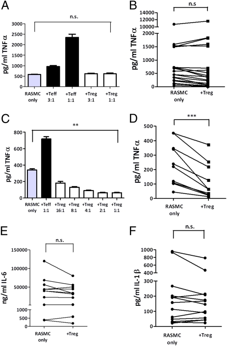
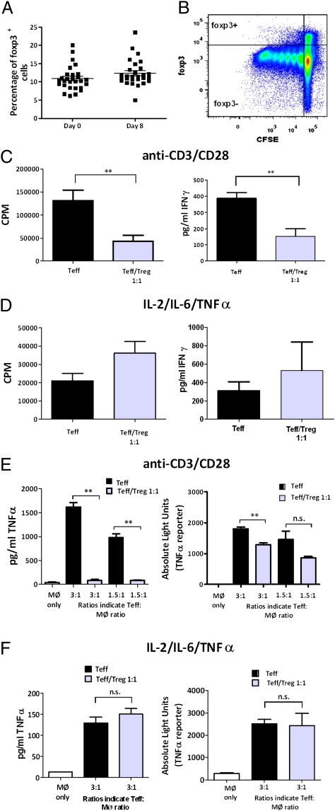
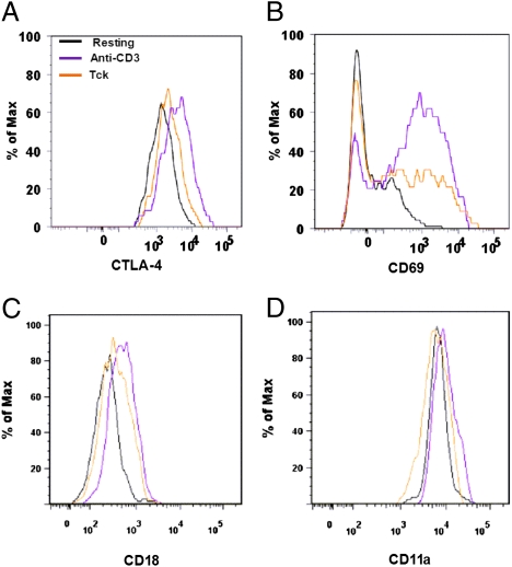
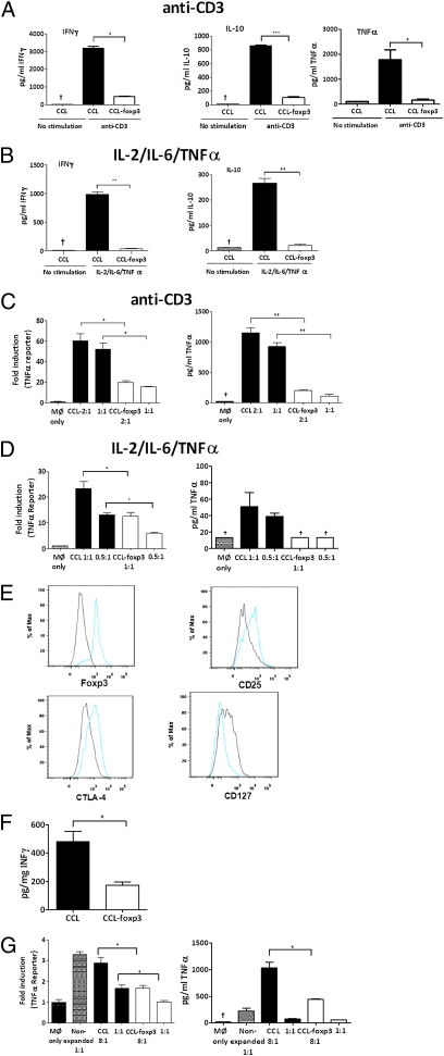
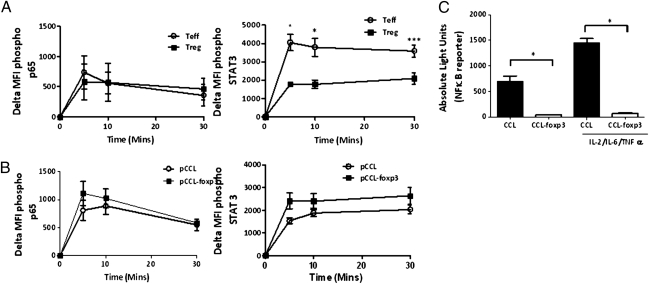
Increasing evidence suggests that regulatory T cell (Treg) function is impaired in chronic inflammatory diseases such as rheumatoid arthritis (RA). Here we demonstrate that Tregs are unable to modulate the spontaneous production of TNF-α from RA synovial cells cultured from the diseased synovium site. Cytokine (IL-2, IL-6, TNF-α) activated T cells (Tck), cells we previously demonstrated to mimic the effector function of pathogenic RA synovial T cells, contained Tregs that survived and divided in this cytokine environment; however, the up-regulation of key molecules associated with Treg function (CTLA-4 and LFA-1) was impaired. Furthermore, Tregs were unable to suppress the function of Tcks, including contact-dependent induction of TNF-α from macrophages, supporting the concept that impaired Treg function/responsiveness contributes to chronicity of RA. However, ectopic foxp3 expression in both Tcks and pathogenic RA synovial T cells attenuated their cytokine production and function, including contact-dependent activation of macrophages. This diminished response to cytokine activation after ectopic foxp3 expression involved inhibited NF-κB activity and differed mechanistically from that displayed endogenously in conventional Tregs. These results suggest that diseases such as RA may perpetuate owing to the inability of Tregs to control cytokine-activated T-cell function. Understanding the mechanism whereby foxp3 attenuates the pathogenic function of synovial T cells may provide insight into the mechanisms of chronicity in inflammatory disease and potentially reveal new therapeutic candidates.


Conflict of interest statement

The authors declare no conflict of interest.

Figures

Fig. 1.
Fig. 1.
RA MNC cytokine production is not modulated by Tregs. CD4+CD25+ blood Tregs from healthy donors were cocultured with RA synovial MNCs with soluble anti-CD3, and supernatants were harvested at 24 h for TNF-α by ELISA. A representative titratable experiment (A) and pooled data (B) (n = 19) are shown; and where RA MNCs spontaneously produced <500 pg/mL of TNF-α a representative titratable experiment (C) and pooled data (D) (n = 9) are illustrated. Similarly IL-6 (E) and IL-1β (F) levels were determined in RA MNC cultures with or without CD4+CD25+ Tregs. IFN-γ was not detectable in supernatants/cell lysates. **P < 0.01, ***P < 0.001. n.s., not significant.
Fig. 2.
Fig. 2.
Tregs do not suppress Teffs in cytokine-activated cultures. (A) Proportion of cells expressing foxp3 (mean ± SEM; n = 28) was determined before and after (8 d) stimulation with the Tck cytokine mixture by flow cytometry. (B) Carboxyfluorescein succinimidyl ester (CFSE) content of foxp3+ and foxp3 populations of CD4+CD45RO+ cells at day 8 (n = 3). CD4+CD45RO+CD25 (Teff) cells were cultured in the presence (gray bars) or absence (black bars) of CD4+CD45RO+CD25+ (Treg) cells at a 1:1 ratio and stimulated with either (C) anti-CD3 (1 μg/mL)/anti-CD28 (2.5 μg/mL) or (D) the Tck cytokine mixture. Proliferation was assessed by 3H-thymidine incorporation at 102–120 h. IFN-γ production was determined after 120 h (anti-CD3/anti-CD28) or 8 d (Tck cytokine mixture) (n > 3). Cells were stimulated for (E) 3 d with anti-CD3/anti-CD28 or (F) 8 d with Tck cytokine mixture, then cocultured for 8 h with autologous macrophages transduced with a TNF-α–luciferase reporter. Representative experiments of TNF-α levels (ELISA) and expression of TNF-α reporter are shown. **P < 0.01. n.s., not significant.
Fig. 3.
Fig. 3.
Cytokine stimulation does not induce fully activated Treg phenotype. CD4+CD45RO+ cells were isolated from PBLs by negative selection (5) and activated with either anti-CD3 for 48 h or Tck cytokine mixture for 8 d and expression of intracellular CTLA-4 (A), CD69 (B), CD18 (LFA-1 β-chain) (C), and CD11a (LFA-1 α-chain) (D) on foxp3hi gated cells determined by flow cytometry (n = 4).
Fig. 4.
Fig. 4.
Ectopic foxp3 expression attenuates CD4+ effector function. CD4+ lymphocytes isolated from either blood of healthy donors (A–D) or RA synovial MNCs (E–G) were transduced with either pCCL or pCCL-foxp3 lentivirus. NGFR+ cells were restimulated with either anti-CD3 (5 μg/mL) for 24 h or the Tck cytokine mixture for 5 d. RA synovial CD4+ lymphocytes were restimulated with the Tck cytokine mixture. Ratios indicate T cell:macrophage proportions. Representative experiments of >n = 3 are shown and illustrate cytokine production from anti-CD3 activated (A) and cytokine-stimulated (B) T lymphocytes. TNF-α reporter gene and TNF-α protein levels are shown for cocultures with anti-CD3 (C) and cytokine-stimulated (D) T lymphocytes. (E) Phenotype of synovial CD4+ lymphocytes was determined after transduction with pCCL (black) or pCCL-foxp3 (blue) lentivirus. (F) IFN-γ production from RA CD4+ lymphocytes and (G) TNF-α reporter gene activity and protein levels from RA CD4+ lymphocyte:macrophage cocultures after transduction with pCCL-foxp3. *P < 0.05, **P < 0.01, ***P < 0.001, cytokine concentration below limits of ELISA.
Fig. 5.
Fig. 5.
Ectopic foxp3 expression attenuates NF-κB transcription. Kinetics of p65 and STAT-3 phosphorylation were determined in Teffs and nTregs (A) and pCCL and pCCL-foxp3 (B) transduced cytokine-stimulated Teffs by flow cytometry (mean ± SEM of > n = 3). (C) CD4+ cells transduced with NF-κB-luciferase reporter lentivirus and pCCL/pCCLfo×p3 were stimulated with the Tck cytokine mixture for 2 h and luciferase activity determined (mean ± SEM, n = 4; *P < 0.05).

References

    1. Gregersen PK, Silver J, Winchester RJ. The shared epitope hypothesis. An approach to understanding the molecular genetics of susceptibility to rheumatoid arthritis. Arthritis Rheum. 1987;30:1205–1213. - PubMed
    1. Wagner UG, Koetz K, Weyand CM, Goronzy JJ. Perturbation of the T cell repertoire in rheumatoid arthritis. Proc Natl Acad Sci USA. 1998;95:14447–14452. - PMC - PubMed
    1. Brennan FM, et al. Heterogeneity of T cell receptor idiotypes in rheumatoid arthritis. Clin Exp Immunol. 1988;73:417–423. - PMC - PubMed
    1. Unutmaz D, Pileri P, Abrignani S. Antigen-independent activation of naive and memory resting T cells by a cytokine combination. J Exp Med. 1994;180:1159–1164. - PMC - PubMed
    1. Brennan FM, et al. Resting CD4+ effector memory T cells are precursors of bystander-activated effectors: A surrogate model of rheumatoid arthritis synovial T-cell function. Arthritis Res Ther. 2008;10:R36. - PMC - PubMed

LinkOut - more resources