. 2011 Oct;121(10):3877-88.
doi: 10.1172/JCI59211. Epub 2011 Sep 19.

Loss of memory B cells during chronic HIV infection is driven by Foxo3a- and TRAIL-mediated apoptosis

Affiliations

Loss of memory B cells during chronic HIV infection is driven by Foxo3a- and TRAIL-mediated apoptosis

Julien van Grevenynghe et al. J Clin Invest. 2011 Oct.

Erratum in

  • J Clin Invest. 2012 Jul 2;122(7):2704

Abstract

Loss of memory B cells occurs from the onset of HIV-1 infection and persists into the chronic stages of infection. Lack of survival of these cells, even in subjects being treated, could primarily be the consequence of an altered local microenvironment induced by HIV infection. In this study we showed that memory B cell survival was significantly decreased in aviremic successfully treated (ST) subjects compared with subjects who control viral load as a result of natural immunity (elite controller [EC]) or with uninfected control (HIV-) subjects. The lower survival levels observed in memory B cells from ST subjects were the result of disrupted IL-2 signaling that led to increased transcriptional activity of Foxo3a and increased expression of its proapoptotic target TRAIL. Notably, memory B cell survival in ST subjects was significantly enhanced by the addition of exogenous IL-2 in a Foxo3a-dependent manner. We further showed that Foxo3a silencing by siRNA resulted in decreased expression of TRAIL and apoptosis levels in memory B cells from ST subjects. Our results thus establish a direct role for Foxo3a/TRAIL signaling in the persistence of memory B cells and provide a mechanism for the reduced survival of memory B cells during HIV infection. This knowledge could be exploited for the development of therapeutic and preventative HIV vaccines.


Figures

Figure 1
Figure 1. Loss of peripheral CD27+ memory B cells in ST subjects is associated with higher levels of apoptosis.
(AC) Ex vivo PBMCs from ST, EC, and HIV subjects were stained with anti-CD3–PB (PB, pacific blue), anti-CD19–Alexa Fluor 700, and anti-CD27–APC Cy7 Abs to measure (A) the percentages of CD19+ B cells within total lymphocytes, (B) the absolute number of CD27+ memory B cells per mm3 of blood in EC and ST subjects using available clinical data for HIV+ subjects (n = 13), and (C) the frequency of the memory population in total B cells (n = 15). (D) Memory B cells (105) from HIV+ and HIV subjects were purified and cocultured for 7 days with 9 × 10⁵ autologous CD19-depleted PBMCs in the presence of 10 μM AZT. The absolute number of viable memory B cells was determined on day 7 using trypan blue exclusion (n = 5). (E) The percentages of apoptotic memory B cells from ST, EC, and HIV subjects on day 7 were analyzed using annexin V–APC staining. The histograms are representative of raw data from 5 independent experiments. (F) The correlation between the absolute numbers of viable memory B cells and the frequency of apoptosis of this population at day 7 of coculture was calculated (n = 15; Spearman test).
Figure 2
Figure 2. Foxo3a regulated the premature death of memory B cells in ST subjects.
Purified memory B cells from ST subjects were either electroporated (electr.), or transfected with negative (neg.) siRNAs or with specific Foxo3a siRNAs, and then cocultured for 7 days with autologous CD19 PBMCs. (A) The efficacy of silencing Foxo3a using siRNAs was determined on purified memory B cells from ST subjects after 2 days of coculture using Western blot analysis. No changes were observed in the levels of total ERK expression, confirming the specificity of Foxo3a silencing. (B) Densitometric quantification of specific bands was performed using ImageQuant software (15, 17). The levels of expression of each protein were normalized to actin and were later expressed as the ratio of densitometric values of protein of interest divided by densitometric values of actin within the same blot. Results shown represent the mean relative expression ± SD of 5 independent experiments. (C) The levels of apoptosis in the presence or absence of Foxo3a were assessed on transfected memory B cells from ST subjects on day 7 by annexin V–APC staining (mean ± SD). The histograms shown are representative of raw data from 5 independent experiments.
Figure 3
Figure 3. Interfering with TRAIL pathway enhances the survival of memory B cells from ST subjects.
(A) Western blot analysis of Bim and TRAIL expression was performed on ex vivo cell lysates of highly purified memory B cells isolated from HIV+ and HIV subjects. (B) Densitometric quantification of 5 independent experiments was performed using ImageQuant software (mean ± SD). (C) Ex vivo PBMCs from HIV+ and HIV subjects were stained with 7AAD, anti-CD3–PB, anti-CD19–Alexa Fluor 700, anti-CD27–APC Cy7, and anti-TRAIL–PE Abs. The levels of surface-bound TRAIL on gated viable memory B cells are shown with their representative histograms (n = 15). Similar results were obtained for MFI. (D) The correlation between the frequency of memory B cells and the surface expression levels of TRAIL on these cells is shown (n = 45; Spearman test). (EG) Memory B cells from ST and HIV subjects were pretreated with TRAILi and then cocultured for 7 days with autologous CD19 PBMCs (n = 5). (E and F) The levels of apoptosis as measured by annexin V staining (E) and the surface expression levels of TRAIL (F) in the presence or absence of TRAILi were assessed on memory B cells on day 7. (G) Correlation between the frequency of apoptotic memory B cells and the surface expression levels of TRAIL on these cells is also shown (n = 10; Spearman test).
Figure 4
Figure 4. Reduced IL-2 signaling resulted in premature memory B cell death in HIV+ subjects.
(A) IL-2 was measured in plasma samples from EC, ST, and HIV subjects using ELISA (n ≥ 11 per group). (B) Correlation between the frequency of ex vivo memory B cell subsets within total B cells and plasma IL-2 levels in all subjects was also determined (n = 41; Spearman test). (C and D) Ex vivo PBMCs from ST, EC, and HIV subjects were stained with 7AAD, anti-CD3–PB, anti-CD19–Alexa Fluor 700, anti-CD27–APC Cy7, and anti-CD25–PE (IL-2 receptor) to monitor their surface expression levels on gated viable memory B cells (n = 15). Results show (C) the frequencies of positive cells and (D) the expression levels of CD25 (MFI). (E) PBMCs from HIV+ and HIV subjects were treated for 15 minutes with 10 ng/ml IL-2, then labeled with 7AAD, anti-CD3–PB, anti-CD19–Alexa Fluor 700, and anti-CD27–APC Cy7 Abs and then subjected to PhosFlow staining with anti-pSTAT5–PE Ab. Results show the levels of pSTAT5 within viable memory B cells (MFI; n = 15). Representative histograms for Figure 3, C and E, are also shown.
Figure 5
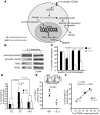
Figure 5. IL-2 treatment of memory B cells from ST subjects led to reduced levels of apoptosis by decreasing TRAIL expression.
(A) Schematic showing the modulation of Foxo3a signaling pathway by IL-2 triggering. (B) Western blot analysis of memory B cells purified from ST subjects untreated or treated with 10 ng/ml of IL-2 for 15 minutes (pFoxo3a) or 24 hours (BimEL and TRAIL) (n = 5). The expression levels of actin were measured on the same blots to control for loading. (C) Densitometric quantification of specific bands was performed using ImageQuant software (mean ± SD). (D and E) Purified memory B cells from ST, EC, and HIV subjects were cocultured for 7 days with autologous CD19 PBMCs in the presence or absence of 10 ng/ml IL-2 (n = 5). UNT, untreated. (D) Percentage of apoptosis ± SD on cocultured memory B cells (after 7 days) for all groups of subjects was determined by annexin V–APC. (E) Surface expression levels of TRAIL on memory B cells from ST subjects in the presence or absence of IL-2 at day 7 of coculture. Histograms shown are representative of 5 different ST subjects. (F) Correlation between surface expression of TRAIL and the percentage of apoptosis in memory B cells from ST subjects at day 7 of coculture in the presence or absence of IL-2 (n = 10; Spearman test).
Figure 6
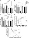
Figure 6. The improvement of ASC survival in ST subjects was achieved by enhancing memory B cell survival.
Purified memory B cells from ST, EC, and HIV subjects were polyclonally activated and cocultured for 3 days in the presence or absence of exogenous IL-2, TRAILi, or Z-VAD. (A) The percentage of apoptosis ± SD was determined on day 3 by annexin V–APC staining (n = 5). (B and C) Memory B cells from HIV+ and HIV subjects were activated for 3 days in ELISPOT plates (n = 5). The plates were then treated with anti-IgG and anti-IgM Abs, followed by the addition of secondary alkaline phosphatase–conjugated Ab to quantify the numbers of (B) IgM- and (C) IgG-secreting cells. Results represent the number of SFCs detected for each condition per 104 purified memory B cells (mean ± SD). Unstimulated memory B cells were used to calculate the nonspecific background. (D) Levels of HIV-specific IgG Abs produced by PBMCs from EC, ST, and HIV subjects activated with PWM for 3 days using ELISA assays. Results showed OD measured at 405 nM. (E) Correlations between the frequency of apoptotic memory B cells and the number of IgG+ ASCs after 3 days of activation are depicted for all subjects (n = 15; Spearman test).
Figure 7
Figure 7. Silencing Foxo3a increased the number of ASCs in ST subjects.
Memory B cells from EC and ST subjects were transfected with Foxo3a siRNA or negative siRNA for 2 days and then polyclonally activated for 3 days. (A) Western blot analysis of Foxo3a and its proapoptotic targets (TRAIL and Bim) was performed on memory B cells from ST subjects after 2 days of transfection and showed reduced Foxo3a, TRAIL, and Bim expression levels. (B) Densitometric values are representative of 5 independent experiments (mean ± SD). (C and D) ELISPOT assay was performed following activation to measure (C) IgM- and (D) IgG-secreting cells as described above. Results are expressed as the mean ASC number ± SD (n = 5).

References

    1. Moir S, Fauci AS. B cells in HIV infection and disease. Nat Rev Immunol. 2009;9(4):235–245. doi: 10.1038/nri2524. - DOI - PMC - PubMed
    1. De Milito A, et al. Mechanisms of hypergammaglobulinemia and impaired antigen-specific humoral immunity in HIV-1 infection. Blood. 2004;103(6):2180–2186. doi: 10.1182/blood-2003-07-2375. - DOI - PubMed
    1. Amadori A, et al. B cell activation during HIV-1 infection. II. Cell-to-cell interactions and cytokine requirement. J Immunol. 1991;146(1):57–62. - PubMed
    1. Samuelsson A, Brostrom C, van Dijk N, Sonnerborg A, Chiodi F. Apoptosis of CD4+ and CD19+ cells during human immunodeficiency virus type 1 infection--correlation with clinical progression, viral load, and loss of humoral immunity. Virology. 1997;238(2):180–188. doi: 10.1006/viro.1997.8790. - DOI - PubMed
    1. Jiang W, et al. Impaired naive and memory B-cell responsiveness to TLR9 stimulation in human immunodeficiency virus infection. J Virol. 2008;82(16):7837–7845. doi: 10.1128/JVI.00660-08. - DOI - PMC - PubMed

Publication types

MeSH terms