. 2011 Sep 18;6(11):714-9.
doi: 10.1038/nnano.2011.151.

Cell entry of one-dimensional nanomaterials occurs by tip recognition and rotation

Affiliations

Cell entry of one-dimensional nanomaterials occurs by tip recognition and rotation

Xinghua Shi et al. Nat Nanotechnol. .

Abstract

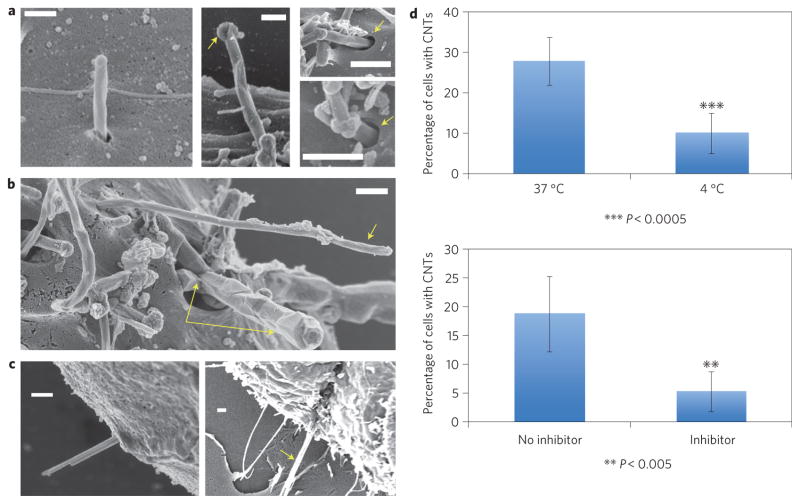
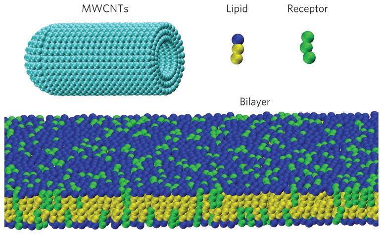
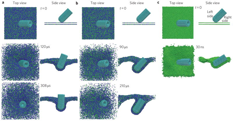
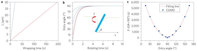
Materials with high aspect ratio, such as carbon nanotubes and asbestos fibres, have been shown to cause length-dependent toxicity in certain cells because these long materials prevent complete ingestion and this frustrates the cell. Biophysical models have been proposed to explain how spheres and elliptical nanostructures enter cells, but one-dimensional nanomaterials have not been examined. Here, we show experimentally and theoretically that cylindrical one-dimensional nanomaterials such as carbon nanotubes enter cells through the tip first. For nanotubes with end caps or carbon shells at their tips, uptake involves tip recognition through receptor binding, rotation that is driven by asymmetric elastic strain at the tube-bilayer interface, and near-vertical entry. The precise angle of entry is governed by the relative timescales for tube rotation and receptor diffusion. Nanotubes without caps or shells on their tips show a different mode of membrane interaction, posing an interesting question as to whether modifying the tips of tubes may help avoid frustrated uptake by cells.


Conflict of interest statement

The authors declare no competing financial interests.

Figures

Figure 1
Figure 1. Experimental evidence for energy-dependent tip-entry mode in the cellular interactions of one-dimensional nanomaterials
ac, Field-emission SEM images following fixation and osmium tetroxide staining. Scale bars, 300 nm. a, Entry of MWCNTs into murine liver cells. Left: MWCNT undergoing high-angle tip entry. Middle: arrow shows carbon shell that distinguishes nanotubes from surface microvilli. Right: arrows show membrane invaginations at the point of entry, characteristic of endocytosis. b, Isolated MWCNT (single arrow) and tube bundle (double arrow) entering human mesothelial cells at high angle. c, Gold nanowires (30 nm, left) and 500 nm crocidolite asbestos fibre (right) entering mesothelial cells through their tips. d, Reduced uptake at 4 °C (top) and in the presence of a metabolic inhibitor mixture (bottom) containing NaF, NaN3 and antimycin A. Error bars show standard error; asterisks indicate that the temperature and inhibitor effects are statistically significant (P < 0.0005 and 0.005, respectively), confirming energy-dependent endocytosis.
Figure 2
Figure 2. Course-grained molecular dynamics simulation model
Models of DPPC lipid and receptor molecules formed by one hydrophilic head-bead and two hydrophobic tail-beads, and a capped MWCNT with diameter d = 20 nm and length L = 46 nm consisting of three concentric walls. A membrane bilayer consisting of lipid and receptor molecules spans the simulation box.
Figure 3
Figure 3. Time sequence of CGMD simulation results showing a MWCNT penetrating the cell membrane at an initial entry angle of θ 0 = 45°
a, At receptor (green) density φ = 0.25, the MWCNT rotates to 90° before being fully wrapped. b, At receptor density φ = 0.33, the tube is fully wrapped before reaching the 90° entry angle. c, At receptor density φ = 1, the tube rotates towards a low entry angle.
Figure 4
Figure 4. Analytical model of a MWCNT entering cell
a,b, Timescales associated with wrapping and tip rotation: entry time as a function of the engulfed length (a) under selected parameter values eRL = 15, B = 20, ξL = 5,000 μm−2, ξ̃ = 0.1, d = 20 nm, D = 1 × 10³ (red line) or 1 × 10⁴ nm2 s−1 (blue line); evolution of the entry angle of the MWCNT (b) at different initial entry angles θ0 = 1°, 15°, 45° and 75°. The other parameters are d = 20 nm, L = Li + Le = 30 μm, ηe = 1 × 10⁻³ Pa s, η i = 10 Pa s. Inset to b: schematic of an MWCNT entering a cell at angle θ. The curved bilayer exerts torque T onto the MWCNT. c, Total elastic energy as a function of the entry angle of the MWCNT relative to its reference value at the entry angle of 90°. The solid red line is a fit from CGMD simulation results (blue squares).

References

    1. Poland CA, et al. Carbon nanotubes introduced into the abdominal cavity of mice show asbestos-like pathogenicity in a pilot study. Nature Nanotech. 2008;3:423–428. - PubMed
    1. Donaldson K, Murphy FA, Duffin R, Poland CA. Asbestos, carbon nanotubes and the pleural mesothelium: a review of the hypothesis regarding the role of long fibre retention in the parietal pleura, inflammation and mesothelioma. Part Fibre Toxicol. 2010;7:5. - PMC - PubMed
    1. Brown DM, et al. An in vitro study of the potential of carbon nanotubes and nanofibres to induce inflammatory mediators and frustrated phagocytosis. Carbon. 2007;45:1743–1756.
    1. Gao HJ, Shi WD, Freund LB. Mechanics of receptor-mediated endocytosis. Proc Natl Acad Sci USA. 2005;102:9469–9474. - PMC - PubMed
    1. Decuzzi P, Ferrari M. The receptor-mediated endocytosis of nonspherical particles. Biophys J. 2008;94:3790–3797. - PMC - PubMed

Publication types