

doi: 10.1038/nsmb.2119.
Crystal structure of a monomeric retroviral protease solved by protein folding game players
Affiliations
- PMID: 21926992
- PMCID: PMC3705907
- DOI: 10.1038/nsmb.2119
Item in Clipboard
Crystal structure of a monomeric retroviral protease solved by protein folding game players
Nat Struct Mol Biol.
.
Erratum in
- Nat Struct Mol Biol. 2012 Mar;19(3):364
Abstract
Following the failure of a wide range of attempts to solve the crystal structure of M-PMV retroviral protease by molecular replacement, we challenged players of the protein folding game Foldit to produce accurate models of the protein. Remarkably, Foldit players were able to generate models of sufficient quality for successful molecular replacement and subsequent structure determination. The refined structure provides new insights for the design of antiretroviral drugs.
Figures
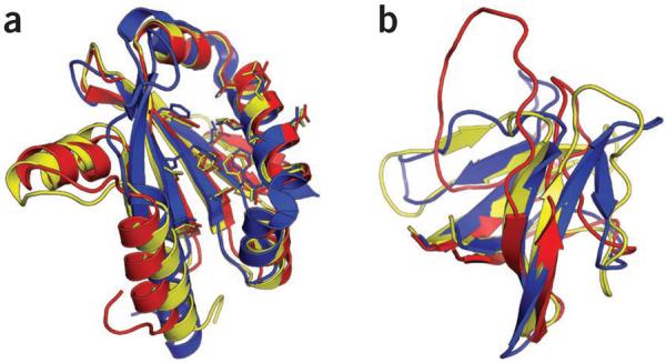
Successful CASP9 predictions by the Foldit Void Crushers Group. (a) Starting from the fourth-ranked Rosetta Server model (red) for CASP9 target T0581, the Foldit Void Crushers Group (yellow) generated a model that was closer to the crystal structure later determined (blue). (b) Starting from a modified Rosetta model built using the Alignment Tool (red), the Foldit Void Crushers Group generated a model (yellow) considerably closer to the later determined crystal structure (blue). Images were produced using PyMOL software (http://www.pymol.org ).

M-PMV retroviral protease structure improvement by the Foldit Contenders Group. (a) Progress of structure refinement over the first 16 d of game play. The x axis shows progression in time, and the y axis shows the Phaser log-likelihood (LLG) of each model in a near-native orientation. To identify a solution as correct by molecular replacement using Phaser, the model must have an LLG better than the best random models. The distribution of these best random predictions is indicated by the intensity of the pale blue band. (Because almost all the models are too poor to allow correct placement in the unit cell, Phaser LLGs are calculated after optimal superposition of each model onto the solved crystal structure and rigid-body optimization.) (b) Starting from a quite inaccurate NMR model (red), Foldit player spvincent generated a model (yellow) considerably more similar to the later determined crystal structure (blue) in the β-strand region. (c) Starting from spvincent's model, Foldit player grabhorn generated a model (magenta) considerably closer to the crystal structure with notable improvement of side-chain conformations in the hydrophobic core. (d) Foldit player mimi made additional improvements (in the loop region at the top left) and generated a model (green) of sufficient accuracy to provide an unambiguous molecular replacement solution which allowed rapid determination of the ultimate crystal structure (blue).
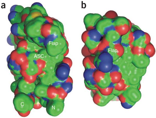
CPK representation of retropepsin surface. (a) The surface of HIV-1 PR protomer extracted from the dimeric molecule (PDB 3hvp), as seen from the direction of the removed dimerization partner. (b) M-PMV PR monomer shown in the same orientation and scale. In this view, the N and C termini (missing in M-PMV PR) are at the bottom, and the flap loop is at the top. The active-site cavity (ASC) is clearly seen between the flap and the body of the HIV-1 PR molecule. In M-PMV PR, the cavity is completely covered by the curled flap.
Comment in
-
Science and Culture: Putting a game face on biomedical research.Proc Natl Acad Sci U S A. 2016 Jun 14;113(24):6577-8. doi: 10.1073/pnas.1607585113. Proc Natl Acad Sci U S A. 2016. PMID: 27302944 Free PMC article. No abstract available.
References
-
- Rohl CA, Strauss CE, Misura KM, Baker D. Methods Enzymol. 2004;383:66–93. - PubMed
Publication types
MeSH terms
Substances
Associated data
- Actions
Grants and funding
LinkOut - more resources
Full Text Sources
Other Literature Sources
