

Ligand discovery from a dopamine D3 receptor homology model and crystal structure
- PMID: 21926995
- PMCID: PMC3197762
- DOI: 10.1038/nchembio.662
Ligand discovery from a dopamine D3 receptor homology model and crystal structure
Abstract
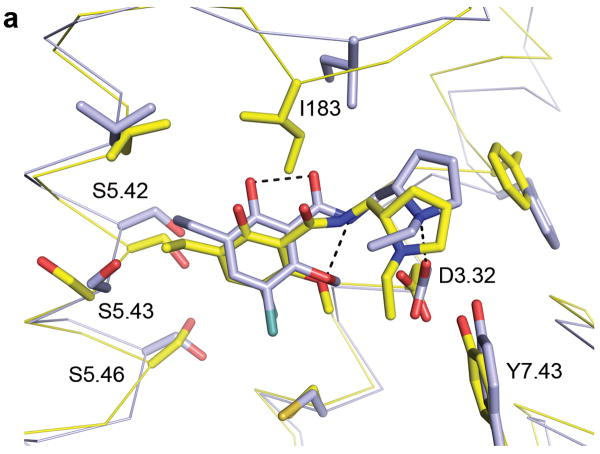
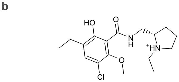
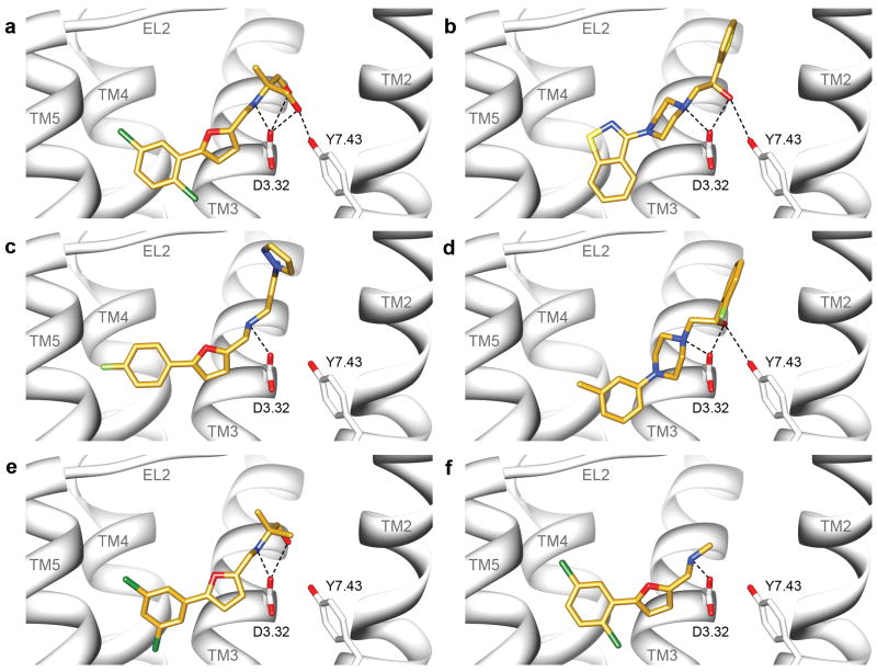
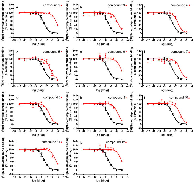
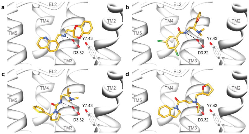
G protein-coupled receptors (GPCRs) are intensely studied as drug targets and for their role in signaling. With the determination of the first crystal structures, interest in structure-based ligand discovery increased. Unfortunately, for most GPCRs no experimental structures are available. The determination of the D(3) receptor structure and the challenge to the community to predict it enabled a fully prospective comparison of ligand discovery from a modeled structure versus that of the subsequently released crystal structure. Over 3.3 million molecules were docked against a homology model, and 26 of the highest ranking were tested for binding. Six had affinities ranging from 0.2 to 3.1 μM. Subsequently, the crystal structure was released and the docking screen repeated. Of the 25 compounds selected, five had affinities ranging from 0.3 to 3.0 μM. One of the new ligands from the homology model screen was optimized for affinity to 81 nM. The feasibility of docking screens against modeled GPCRs more generally is considered.
Figures
References
-
- Overington JP, Al-Lazikani B, Hopkins AL. How many drug targets are there? Nat Rev Drug Discov. 2006;5:993–996. - PubMed
Publication types
MeSH terms
Substances
Associated data
- PubChem-Substance/125092008
- PubChem-Substance/125092009
- PubChem-Substance/125092010
- PubChem-Substance/125092011
- PubChem-Substance/125092012
- PubChem-Substance/125092013
- PubChem-Substance/125092014
- PubChem-Substance/125092015
- PubChem-Substance/125092016
- PubChem-Substance/125092017
- PubChem-Substance/125092018
- PubChem-Substance/125092019
- PubChem-Substance/125092020
- PubChem-Substance/125092021
- PubChem-Substance/125092022
- PubChem-Substance/125092023
- PubChem-Substance/125092024
- PubChem-Substance/125092025
- PubChem-Substance/125092026
- PubChem-Substance/125092027
- PubChem-Substance/125092028
- PubChem-Substance/125092029
- PubChem-Substance/125092030
- PubChem-Substance/125092031
- PubChem-Substance/125092032
- PubChem-Substance/125092033
- PubChem-Substance/125092034
- PubChem-Substance/125092035
- PubChem-Substance/125092036
- PubChem-Substance/125092037
- PubChem-Substance/125092038
- PubChem-Substance/125092039
- PubChem-Substance/125092040
- PubChem-Substance/125092041
- PubChem-Substance/125092042
- PubChem-Substance/125092043
- PubChem-Substance/125092063
- PubChem-Substance/125092064
- PubChem-Substance/125092065
- PubChem-Substance/125092066
- PubChem-Substance/125092067
- PubChem-Substance/125092068
- PubChem-Substance/125092069
- PubChem-Substance/125092070
- PubChem-Substance/125092071
- PubChem-Substance/125092072
- PubChem-Substance/125092073
- PubChem-Substance/125092074
- PubChem-Substance/125092075
- PubChem-Substance/125092076
- PubChem-Substance/125092077
- PubChem-Substance/125092078
- PubChem-Substance/125092079
- PubChem-Substance/125092080
- PubChem-Substance/125092081
- PubChem-Substance/125092082
- PubChem-Substance/125092083
- PubChem-Substance/125092084
- PubChem-Substance/125092085
- PubChem-Substance/125092086
- PubChem-Substance/125092087
- PubChem-Substance/125092088
- PubChem-Substance/125092089
- PubChem-Substance/125092090
- PubChem-Substance/125092091
- PubChem-Substance/125092092
- PubChem-Substance/125092093
- PubChem-Substance/125092094
- PubChem-Substance/125092095
- PubChem-Substance/125092096
- PubChem-Substance/125092097
- PubChem-Substance/125092098
Grants and funding
- U19 MH082441/MH/NIMH NIH HHS/United States
- R01 GM071630/GM/NIGMS NIH HHS/United States
- GM59957/GM/NIGMS NIH HHS/United States
- R01 DA017204/DA/NIDA NIH HHS/United States
- F32 GM088991/GM/NIGMS NIH HHS/United States
- F32GM088991/GM/NIGMS NIH HHS/United States
- R01GM54762/GM/NIGMS NIH HHS/United States
- GM71630/GM/NIGMS NIH HHS/United States
- U54GM093342/GM/NIGMS NIH HHS/United States
- R01 DA027170/DA/NIDA NIH HHS/United States
- F32 GM096544/GM/NIGMS NIH HHS/United States
- F32GM096544/GM/NIGMS NIH HHS/United States
- R01 GM054762/GM/NIGMS NIH HHS/United States
- GM71790/GM/NIGMS NIH HHS/United States
- U54 GM093342/GM/NIGMS NIH HHS/United States
- R01 GM059957/GM/NIGMS NIH HHS/United States
LinkOut - more resources
Full Text Sources
Other Literature Sources
Chemical Information
