. 2011 Sep 18;7(11):779-86.
doi: 10.1038/nchembio.657.

RF1 knockout allows ribosomal incorporation of unnatural amino acids at multiple sites

Affiliations

RF1 knockout allows ribosomal incorporation of unnatural amino acids at multiple sites

David B F Johnson et al. Nat Chem Biol. .

Abstract

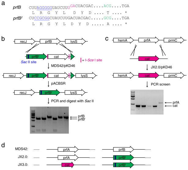
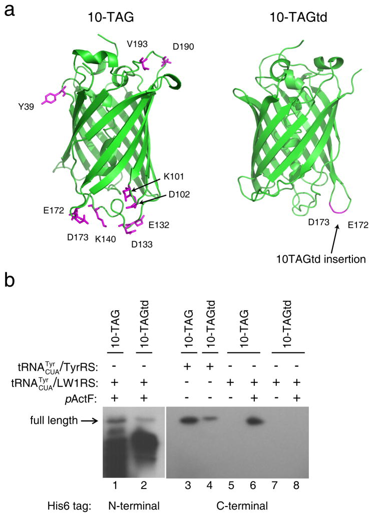
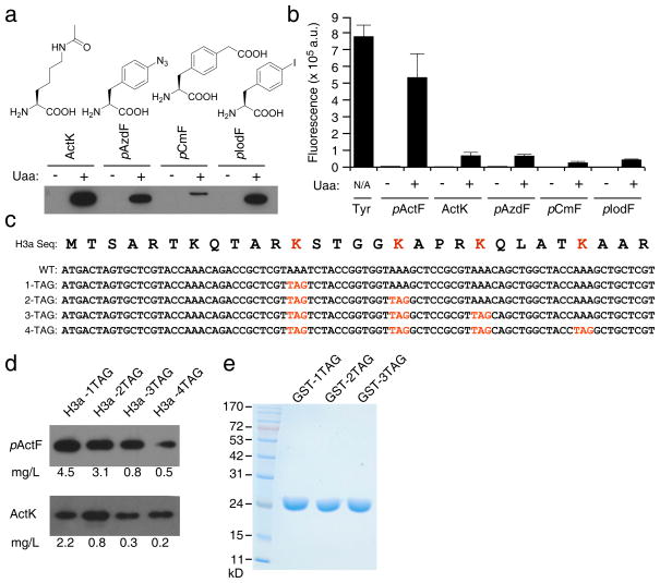
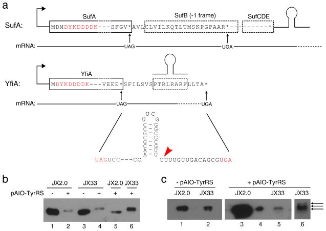
Stop codons have been exploited for genetic incorporation of unnatural amino acids (Uaas) in live cells, but their low incorporation efficiency, which is possibly due to competition from release factors, limits the power and scope of this technology. Here we show that the reportedly essential release factor 1 (RF1) can be knocked out from Escherichia coli by 'fixing' release factor 2 (RF2). The resultant strain JX33 is stable and independent, and it allows UAG to be reassigned from a stop signal to an amino acid when a UAG-decoding tRNA-synthetase pair is introduced. Uaas were efficiently incorporated at multiple UAG sites in the same gene without translational termination in JX33. We also found that amino acid incorporation at endogenous UAG codons is dependent on RF1 and mRNA context, which explains why E. coli tolerates apparent global suppression of UAG. JX33 affords a unique autonomous host for synthesizing and evolving new protein functions by enabling Uaa incorporation at multiple sites.


Conflict of interest statement

Competing financial interests

The authors declare no competing financial interests.

Figures

Figure 1
Figure 1. RF1 can be knocked out from E. coli after RF2 is fixed
(a) Features of the RF2-encoding prfB gene in K-12 E. coli strains: an in-frame UGA stop codon (magenta) for autoregulation of RF2 expression and the Ala246Thr mutation (green) impairing RF2’s release activity for the UAA codon. In prfBf, the UGA regulation was removed and residue 246 reverted to Ala. A Shine-Dalgarno like sequence (blue) in prfB was silently mutated to a Sac II site (blue) in prfBf to facilitate the screening of prfBf knock-in. (b) Generation of the JX2.0 strain. The prfB gene in MDS42 was first replaced with prfBf followed by a Cm resistant cat cassette. The cat cassette was subsequently removed by pACBSR (see Supplementary Methods). (c) Generation of the JX3.0 strain. The RF1-encoding prfA gene was successfully knocked out with a cat cassette in JX2.0 to afford JX3.0. (d) Illustration of main features of the three strains, MDS42, JX2.0 and JX3.0.
Figure 2
Figure 2. RF1 knockout enables incorporation of natural or unnatural amino acids at multiple UAG sites in JX33
(a) Features of the All-in-One plasmid and structures of Tyr and pActF. (b) Western analysis of EGFP expression when UAG codons were decoded as Tyr by pAIO-TyrRS. The same number of cells were used for each sample, and the blot was probed with a penta-His antibody. (c) In-cell fluorescence assay of EGFP intensity when UAG codons were decoded as Tyr by pAIO-TyrRS. The same number of cells was used for each sample. Measurement was performed on 3 independent batches of cells and error bars represent s.e.m.. (d) Western analysis of EGFP expression when UAG codons were decoded as the Uaa pActF by pAIO-LW1RS. Conditions are the same in (b). For each sample, a duplicate of cultures were grown in the presence or absence of pActF in the growth media. (e) In-cell fluorescence assay of EGFP intensity when UAG codons were decoded as pActF by pAIO-LW1RS. Measurements were performed as in (c), n = 3, error bars represent s.e.m. (f) Fluorescence images of cells when UAG was decoded as Tyr or pActF. (g) SDS-PAGE analysis of EGFP proteins with pActF incorporated in JX33 or in BL21(DE3) coexpressing plasmid pET-L11C. The same pAIO-LW1RS plasmids containing 1- or 3-TAG EGFP mutant gene were used in both cells. EGFP was purified with Ni-NTA chromatography. Loading was normalized to the same number of cells, and the gel was stained with Coomassie blue.
Figure 3
Figure 3. Mass spectrometric analyses of EGFP expressed in JX33 show that pActF was selectively incorporated at multiple UAG sites with high fidelity
(a) MS/MS spectrum of EGFP peptide ADHUQQNTPIGDGPVLLPDNHY. U represents the UAG codon at residue 182. Star (*) in the spectrum denotes peptide fragments containing pActF, which unambiguously indicate that pActF was incorporated at the UAG site. (b) Extracted ion chromatograms (EIC) of the above peptide containing pActF (top) or Gln (bottom) at the UAG 182 position. The peak areas are indicated.
Figure 4
Figure 4. Ten UAG sites are simultaneously suppressed with natural or unnatural amino acids in JX33
(a) GFP structure (PDB 1GFL) illustrating the sites where 10 UAG codons were introduced. (b) Western blot analysis of the expression of 10-TAG and 10-TAGtd EGFP in JX33. The UAG codons were decoded as Tyr by tRNACUATyr/TyrRS or as pActF by tRNACUATyr/LW1RS. Cell lysates from same number of cells were separated by SDS-PAGE and probe with a penta-His antibody.
Figure 5
Figure 5. JX33 enables multisite incorporation of various Uaas and in different proteins
(a) Western analysis of the expression of 3-TAG EGFP reporter in JX33 with the UAG codon decoded as different Uaas. The same number of cells was used for each sample. After cell lysis, proteins were separated on SDS-PAGE and probed with a penta-His antibody. Densitometric analysis of Western blot bands and purified protein yields (Supplementary Table 1) were consistent on incorporation efficiency. (b) In-cell fluorescence assay of these mutant EGFP proteins containing different Uaas at 3 UAG sites. Measurements were performed using 3 independent batches of cells. Error bars represent s.e.m.. (c) The N-terminal sequence of human histone H3a and UAG codons introduced at the known acetylation sites. (d) Western analysis showing that full-length histone H3a was expressed for 1-, 2-, 3-, and 4-TAG H3a constructs in the presence of pActF or ActK in JX33 cells. For pActF, the sample loading ratio was 1:1:3:3; for ActK, the sample loading ratio was 1:3:7:9. Yields of H3a proteins were calculated after purification and shown at the bottom. (e) SDS-PAGE analysis of GST proteins expressed in JX33 with pActF incorporated at 1, 2, and 3 UAG sites. GST was purified with Ni-NTA chromatography, separated on the gel and stained with Coomassie blue. Loading was normalized to the same number of cells. Measurements of GST yields were performed on 3 independent batches of cells and errors represent s.e.m..
Figure 6
Figure 6. Legitimate UAG codons of endogenous genes are suppressed efficiently after RF1 knockout, and protein extension beyond the UAG codon is dependent on mRNA context
(a) Two categories of endogenous genes ending in TAG: sufA representing those with an in-frame, non-UAG stop codon downstream before a transcription terminator (shown as a hairpin); yfiA representing those with a terminator before the next in-frame, non-UAG stop codon. A FLAG-tag (DYKDDDDK) was inserted scarlessly to the N-terminus of these two proteins in the genome to facilitate protein detection. Red arrow indicates the end of the mRNA as defined by the terminator. (b) Western analysis of SufA purified from cells. Extension of SufA to its next stop codon occurred only in lanes 4 and 6; all other lanes showed a band corresponding to the wild type SufA. Loading was normalized to the same number of cells in lane 1–4. Lane 5 and 6 were duplicates of lane 2 and 4, respectively, with increasing amounts. An anti-FLAG antibody was used for detection. (c) Western analysis of YfiA purified from cells. Loading in lane 1, 2, 3 and 5 were normalized to the same number of cells. Loading in lane 4 is 1/50 of lane 3, which showed it is a single band. Lane 6 is a re-run of lane 5 sample in a higher percentage gel (20% vs. 15%) for longer time to achieve better separation. Lane 5 and 6 showed 3 predominant bands, while other samples showed only one band.

References

    1. Osawa S, Jukes T, Watanabe K, Muto A. Recent evidence for evolution of the genetic code. Microbiol Rev. 1992;56:229–64. - PMC - PubMed
    1. Knight RD, Freeland SJ, Landweber LF. Rewiring the keyboard: evolvability of the genetic code. Nat Rev Genet. 2001;2:49–58. - PubMed
    1. Benzer S, Champe SP. A change from nonsense to sense in the genetic code. Proc Natl Acad Sci U S A. 1962;48:1114–21. - PMC - PubMed
    1. Beier H, Grimm M. Misreading of termination codons in eukaryotes by natural nonsense suppressor tRNAs. Nucleic Acids Res. 2001;29:4767–82. - PMC - PubMed
    1. Wang L, Brock A, Herberich B, Schultz PG. Expanding the genetic code of Escherichia coli. Science. 2001;292:498–500. - PubMed

Publication types

MeSH terms